INRAE, Avignon Université, UMR SQPOV, Avignon, France.
Instituto de Tecnologia Quimica e Biologica, Universidade Nova de Lisboa, Oeiras, Portugal.
mSphere. 2021 Apr 21;6(2):e00007-21. doi: 10.1128/mSphere.00007-21.
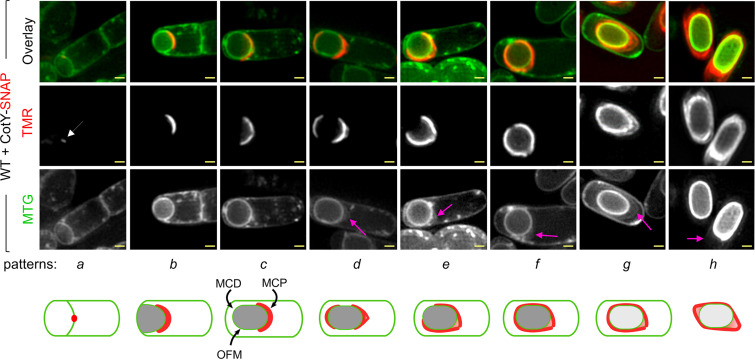
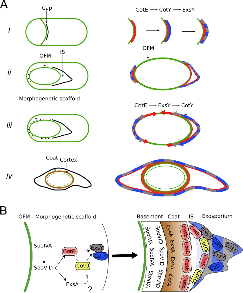
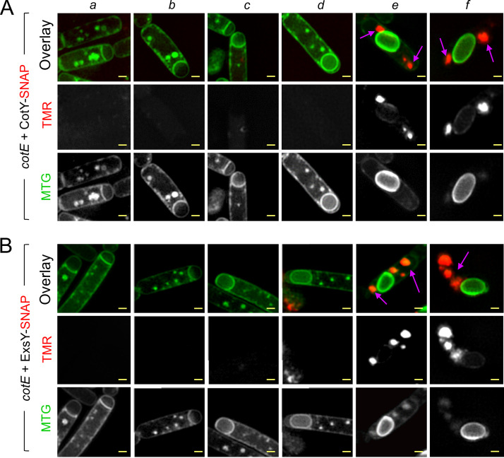
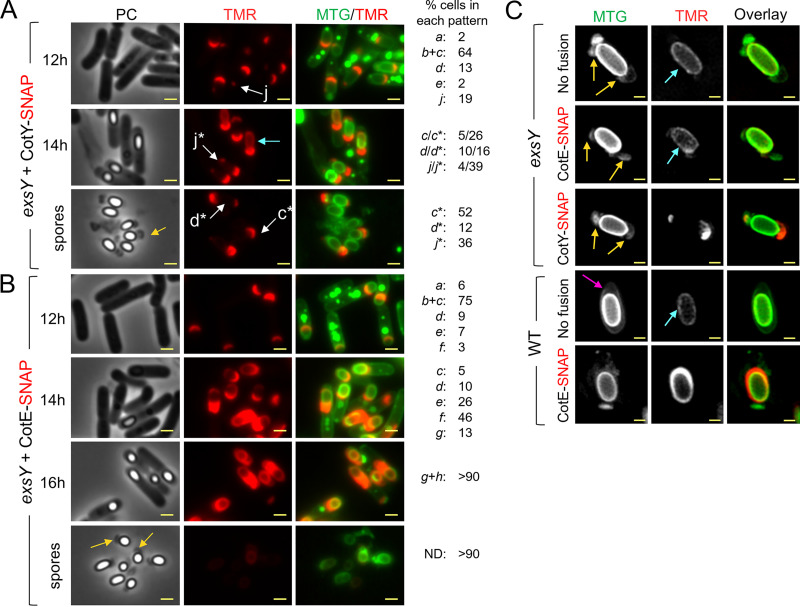
The exosporium is the outermost spore layer of some and species and related organisms. It mediates the interactions of spores with their environment, modulates spore adhesion and germination, and has been implicated in pathogenesis. In , the exosporium consists of a crystalline basal layer, formed mainly by the two cysteine-rich proteins CotY and ExsY, surrounded by a hairy nap composed of glycoproteins. The morphogenetic protein CotE is necessary for the integrity of the exosporium, but how CotE directs exosporium assembly remains unknown. Here, we used super-resolution fluorescence microscopy to follow the localization of SNAP-tagged CotE, CotY, and ExsY during sporulation and evidenced the interdependencies among these proteins. Complexes of CotE, CotY, and ExsY are present at all sporulation stages, and the three proteins follow similar localization patterns during endospore formation that are reminiscent of the localization pattern of CotE. We show that CotE guides the formation of one cap at both forespore poles by positioning CotY and then guides forespore encasement by ExsY, thereby promoting exosporium elongation. By these two actions, CotE ensures the formation of a complete exosporium. Importantly, we demonstrate that the assembly of the exosporium is not a unidirectional process, as previously proposed, but occurs through the formation of two caps, as observed during coat morphogenesis, suggesting that a general principle governs the assembly of the spore surface layers of Spores of are enveloped in an outermost glycoprotein layer. In the group, encompassing the and pathogens, this layer is easily recognizable by a characteristic balloon-like appearance and separation from the underlying coat by an interspace. In spite of its importance for the environmental interactions of spores, including those with host cells, the mechanism of assembly of the exosporium is poorly understood. We used super-resolution fluorescence microscopy to directly visualize the formation of the exosporium during the sporulation of , and we studied the localization and interdependencies of proteins essential for exosporium morphogenesis. We discovered that these proteins form a morphogenetic scaffold before a complete exosporium or coat is detectable. We describe how the different proteins localize to the scaffold and how they subsequently assemble around the spore, and we present a model for the assembly of the exosporium.
外壁是某些 和 物种及相关生物的最外层孢子层。它介导孢子与环境的相互作用,调节孢子的附着和萌发,并与发病机制有关。在 中,外壁由主要由两个富含半胱氨酸的蛋白质 CotY 和 ExsY 形成的结晶基底层组成,周围是由糖蛋白组成的毛茸茸的绒毛。形态发生蛋白 CotE 是外壁完整性所必需的,但 CotE 如何指导外壁组装仍不清楚。在这里,我们使用超分辨率荧光显微镜跟踪 SNAP 标记的 CotE、CotY 和 ExsY 在 孢子形成过程中的定位,并证明了这些蛋白质之间的相互依赖性。CotE、CotY 和 ExsY 的复合物存在于所有孢子形成阶段,并且这三种蛋白质在形成内生孢子的过程中表现出相似的定位模式,这让人联想到 CotE 的定位模式。我们表明,CotE 通过定位 CotY 在两个前孢子极处形成一个帽,然后通过 ExsY 引导前孢子包被,从而促进外壁伸长。通过这两个动作,CotE 确保了完整外壁的形成。重要的是,我们证明了外壁的组装不是如前所述的单向过程,而是通过形成两个帽来进行的,就像在 外壳形态发生过程中观察到的那样,这表明一个普遍的原则支配着 孢子表面层的组装。 的孢子被最外层的糖蛋白层包裹。在 群中,包括 和 病原体,这个层通过一个间隙与下面的外壳分离,很容易识别出其特征气球状外观。尽管外壁对于孢子与环境的相互作用很重要,包括与宿主细胞的相互作用,但外壁的组装机制仍知之甚少。我们使用超分辨率荧光显微镜直接观察 在孢子形成过程中外壁的形成,并研究了对外壁形态发生至关重要的蛋白质的定位和相互依赖性。我们发现,这些蛋白质在可检测到完整的外壁或外壳之前形成一个形态发生支架。我们描述了不同的蛋白质如何定位到支架上,以及它们如何随后围绕孢子组装,并提出了一个外壁组装的模型。