• 文献检索
  • 文档翻译
  • 深度研究
  • 学术资讯
  • Suppr Zotero 插件Zotero 插件
  • 邀请有礼
  • 套餐&价格
  • 历史记录
应用&插件
Suppr Zotero 插件Zotero 插件浏览器插件Mac 客户端Windows 客户端微信小程序
定价
高级版会员购买积分包购买API积分包
服务
文献检索文档翻译深度研究API 文档MCP 服务
关于我们
关于 Suppr公司介绍联系我们用户协议隐私条款
关注我们

Suppr 超能文献

核心技术专利:CN118964589B侵权必究
粤ICP备2023148730 号-1Suppr @ 2026

文献检索

告别复杂PubMed语法,用中文像聊天一样搜索,搜遍4000万医学文献。AI智能推荐,让科研检索更轻松。

立即免费搜索

文件翻译

保留排版,准确专业,支持PDF/Word/PPT等文件格式,支持 12+语言互译。

免费翻译文档

深度研究

AI帮你快速写综述,25分钟生成高质量综述,智能提取关键信息,辅助科研写作。

立即免费体验

眼底照相和光相干断层扫描的野生患者特定信息评估。

Assessment of patient specific information in the wild on fundus photography and optical coherence tomography.

机构信息

Department of Ophthalmology, Inselspital, Bern University Hospital, Bern, Switzerland.

Bern Photographic Reading Center (BPRC), Bern University Hospital, Inselspital, Bern, Switzerland.

出版信息

Sci Rep. 2021 Apr 21;11(1):8621. doi: 10.1038/s41598-021-86577-5.

DOI:10.1038/s41598-021-86577-5
PMID:33883573
原文链接:https://pmc.ncbi.nlm.nih.gov/articles/PMC8060417/
Abstract

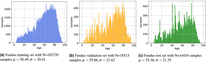
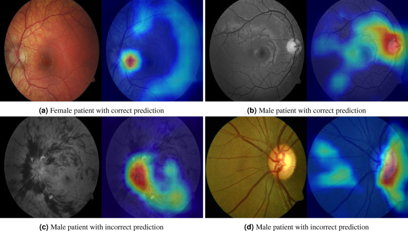
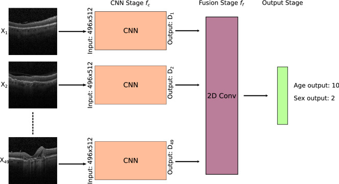
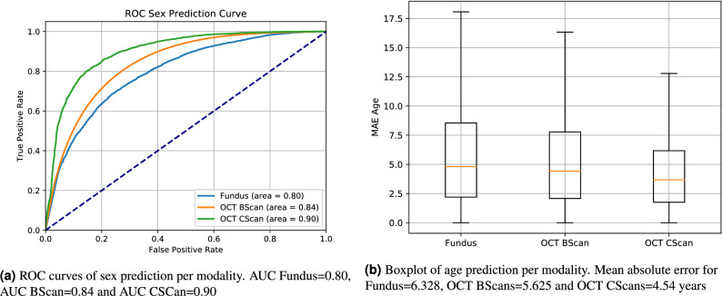
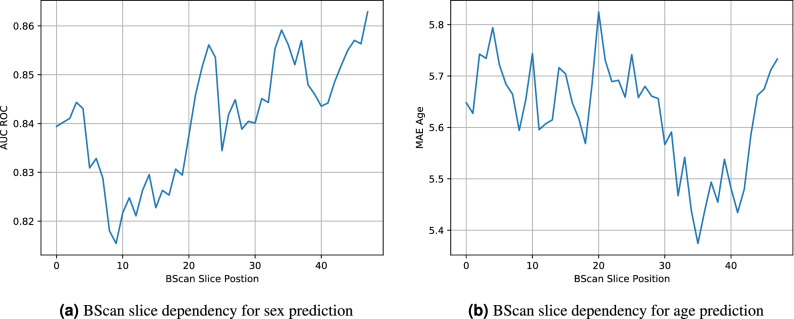
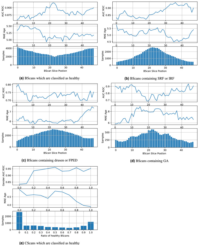
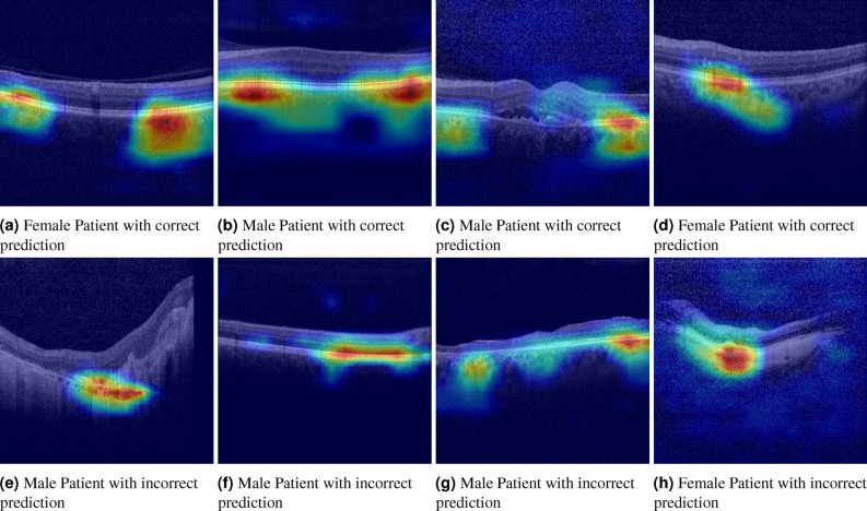
In this paper we analyse the performance of machine learning methods in predicting patient information such as age or sex solely from retinal imaging modalities in a heterogeneous clinical population. Our dataset consists of N = 135,667 fundus images and N = 85,536 volumetric OCT scans. Deep learning models were trained to predict the patient's age and sex from fundus images, OCT cross sections and OCT volumes. For sex prediction, a ROC AUC of 0.80 was achieved for fundus images, 0.84 for OCT cross sections and 0.90 for OCT volumes. Age prediction mean absolute errors of 6.328 years for fundus, 5.625 years for OCT cross sections and 4.541 for OCT volumes were observed. We assess the performance of OCT scans containing different biomarkers and note a peak performance of AUC = 0.88 for OCT cross sections and 0.95 for volumes when there is no pathology on scans. Performance drops in case of drusen, fibrovascular pigment epitheliuum detachment and geographic atrophy present. We conclude that deep learning based methods are capable of classifying the patient's sex and age from color fundus photography and OCT for a broad spectrum of patients irrespective of underlying disease or image quality. Non-random sex prediction using fundus images seems only possible if the eye fovea and optic disc are visible.

摘要

在本文中,我们分析了机器学习方法在从异质临床人群的视网膜成像模式中仅预测患者信息(如年龄或性别)方面的性能。我们的数据集包括 N = 135667 张眼底图像和 N = 85536 张容积 OCT 扫描。深度学习模型被训练用于从眼底图像、OCT 横断面和 OCT 体积预测患者的年龄和性别。对于性别预测,眼底图像的 ROC AUC 为 0.80,OCT 横断面为 0.84,OCT 体积为 0.90。眼底图像的年龄预测平均绝对误差为 6.328 岁,OCT 横断面为 5.625 岁,OCT 体积为 4.541 岁。我们评估了包含不同生物标志物的 OCT 扫描的性能,并注意到在扫描无病变时,OCT 横断面的 AUC 峰值为 0.88,体积的 AUC 峰值为 0.95。在存在 drusen、fibrovascular pigment epitheliuum 脱离和地理萎缩的情况下,性能会下降。我们得出结论,基于深度学习的方法能够从彩色眼底摄影和 OCT 对广泛的患者进行分类,无论潜在疾病或图像质量如何。如果眼底的黄斑和视盘不可见,则似乎只能使用眼底图像进行非随机性别预测。

https://cdn.ncbi.nlm.nih.gov/pmc/blobs/1beb/8060417/20f6b64fb077/41598_2021_86577_Fig9_HTML.jpg
https://cdn.ncbi.nlm.nih.gov/pmc/blobs/1beb/8060417/46c3dbb178f3/41598_2021_86577_Fig1_HTML.jpg
https://cdn.ncbi.nlm.nih.gov/pmc/blobs/1beb/8060417/257103e6446e/41598_2021_86577_Fig2_HTML.jpg
https://cdn.ncbi.nlm.nih.gov/pmc/blobs/1beb/8060417/2e6aaff7b22b/41598_2021_86577_Fig3_HTML.jpg
https://cdn.ncbi.nlm.nih.gov/pmc/blobs/1beb/8060417/04ab1c61a98d/41598_2021_86577_Fig4_HTML.jpg
https://cdn.ncbi.nlm.nih.gov/pmc/blobs/1beb/8060417/4c71061fa5bd/41598_2021_86577_Fig5_HTML.jpg
https://cdn.ncbi.nlm.nih.gov/pmc/blobs/1beb/8060417/5dfe6af7d04b/41598_2021_86577_Fig6_HTML.jpg
https://cdn.ncbi.nlm.nih.gov/pmc/blobs/1beb/8060417/491f2ce6a5b7/41598_2021_86577_Fig7_HTML.jpg
https://cdn.ncbi.nlm.nih.gov/pmc/blobs/1beb/8060417/e3ab0baa3040/41598_2021_86577_Fig8_HTML.jpg
https://cdn.ncbi.nlm.nih.gov/pmc/blobs/1beb/8060417/20f6b64fb077/41598_2021_86577_Fig9_HTML.jpg

相似文献

1
Assessment of patient specific information in the wild on fundus photography and optical coherence tomography.眼底照相和光相干断层扫描的野生患者特定信息评估。
Sci Rep. 2021 Apr 21;11(1):8621. doi: 10.1038/s41598-021-86577-5.
2
The retinal disease screening study: retrospective comparison of nonmydriatic fundus photography and three-dimensional optical coherence tomography for detection of retinal irregularities.视网膜疾病筛查研究:非散瞳眼底照相术与三维光相干断层扫描检测视网膜不规则性的回顾性比较。
Invest Ophthalmol Vis Sci. 2013 Aug 21;54(8):5694-700. doi: 10.1167/iovs.13-12043.
3
Comparison of drusen area detected by spectral domain optical coherence tomography and color fundus imaging.比较频域光相干断层扫描和眼底彩色成像检测到的玻璃膜疣面积。
Invest Ophthalmol Vis Sci. 2013 Apr 3;54(4):2429-34. doi: 10.1167/iovs.12-11569.
4
Comparison of optic area measurement using fundus photography and optical coherence tomography between optic nerve head drusen and control subjects.比较视神经乳头盘状隆起和对照组的眼底照相和光学相干断层扫描的视盘面积测量。
Ophthalmic Physiol Opt. 2013 Mar;33(2):164-71. doi: 10.1111/opo.12017. Epub 2013 Jan 12.
5
Deep Ensemble Learning Based Objective Grading of Macular Edema by Extracting Clinically Significant Findings from Fused Retinal Imaging Modalities.基于深度集成学习的融合视网膜成像模态中临床显著发现提取的黄斑水肿客观分级。
Sensors (Basel). 2019 Jul 5;19(13):2970. doi: 10.3390/s19132970.
6
Deep Learning Models for the Screening of Cognitive Impairment Using Multimodal Fundus Images.深度学习模型在多模态眼底图像认知障碍筛查中的应用。
Ophthalmol Retina. 2024 Jul;8(7):666-677. doi: 10.1016/j.oret.2024.01.019. Epub 2024 Jan 26.
7
From Machine to Machine: An OCT-Trained Deep Learning Algorithm for Objective Quantification of Glaucomatous Damage in Fundus Photographs.从机器到机器:一种基于 OCT 训练的深度学习算法,用于客观量化眼底照片中的青光眼损伤。
Ophthalmology. 2019 Apr;126(4):513-521. doi: 10.1016/j.ophtha.2018.12.033. Epub 2018 Dec 20.
8
Comparison of Color Fundus Photography, Infrared Fundus Photography, and Optical Coherence Tomography in Detecting Retinal Hamartoma in Patients with Tuberous Sclerosis Complex.彩色眼底照相、红外眼底照相和光学相干断层扫描在检测结节性硬化症患者视网膜错构瘤中的比较
Chin Med J (Engl). 2016 May 20;129(10):1229-35. doi: 10.4103/0366-6999.181976.
9
The retinal disease screening study: prospective comparison of nonmydriatic fundus photography and optical coherence tomography for detection of retinal irregularities.视网膜疾病筛查研究:非散瞳眼底照相术与光学相干断层扫描对视盘不规则性检测的前瞻性比较
Invest Ophthalmol Vis Sci. 2013 Feb 19;54(2):1460-8. doi: 10.1167/iovs.12-10727.
10
Sensitivity and Specificity of Potential Diagnostic Features Detected Using Fundus Photography, Optical Coherence Tomography, and Fluorescein Angiography for Polypoidal Choroidal Vasculopathy.眼底照相、光学相干断层扫描和荧光素血管造影检测息肉样脉络膜血管病变的潜在诊断特征的敏感性和特异性。
JAMA Ophthalmol. 2019 Jun 1;137(6):661-667. doi: 10.1001/jamaophthalmol.2019.0565.

引用本文的文献

1
A Deep Learning Model for Detecting the Eyes Receiving Glaucoma Medications Using Anterior Segment Images.一种使用眼前节图像检测接受青光眼药物治疗眼睛的深度学习模型。
Transl Vis Sci Technol. 2025 Aug 1;14(8):28. doi: 10.1167/tvst.14.8.28.
2
Individualized Estimation of Baseline Retinal Nerve Fiber Layer Thickness Using Conditional Variational Autoencoder.使用条件变分自编码器对视网膜神经纤维层厚度基线进行个体化估计。
Ophthalmol Sci. 2025 Jun 9;5(6):100849. doi: 10.1016/j.xops.2025.100849. eCollection 2025 Nov-Dec.
3
Predicting renal function using fundus photography: role of confounders.

本文引用的文献

1
Deep Learning Algorithms and the Protection of Data Privacy.深度学习算法与数据隐私保护
JAMA Ophthalmol. 2020 Oct 1;138(10):1024-1025. doi: 10.1001/jamaophthalmol.2020.2766.
2
Age and sex affect deep learning prediction of cardiometabolic risk factors from retinal images.年龄和性别会影响基于视网膜图像的心血管代谢危险因素的深度学习预测。
Sci Rep. 2020 Jun 10;10(1):9432. doi: 10.1038/s41598-020-65794-4.
3
Effects of Hypertension, Diabetes, and Smoking on Age and Sex Prediction from Retinal Fundus Images.高血压、糖尿病和吸烟对视网膜眼底图像年龄和性别预测的影响。
利用眼底摄影预测肾功能:混杂因素的作用。
Korean J Intern Med. 2025 Mar;40(2):310-320. doi: 10.3904/kjim.2024.076. Epub 2025 Mar 1.
4
Ensemble probabilistic quantization encoding for information preservation of numerical variables in convolutional neural networks.用于卷积神经网络中数值变量信息保留的集成概率量化编码
Sci Rep. 2025 Feb 11;15(1):5021. doi: 10.1038/s41598-025-89430-1.
5
Performance of automated machine learning in detecting fundus diseases based on ophthalmologic B-scan ultrasound images.基于眼科B超超声图像的自动机器学习在检测眼底疾病中的性能。
BMJ Open Ophthalmol. 2024 Dec 11;9(1):e001873. doi: 10.1136/bmjophth-2024-001873.
6
Artificial intelligence-based prediction of neurocardiovascular risk score from retinal swept-source optical coherence tomography-angiography.基于人工智能的视网膜扫频源光相干断层血管造影术预测神经心血管风险评分。
Sci Rep. 2024 Nov 7;14(1):27089. doi: 10.1038/s41598-024-78587-w.
7
Recognition of Patient Gender: A Machine Learning Preliminary Analysis Using Heart Sounds from Children and Adolescents.患者性别的识别:使用儿童和青少年心音的机器学习初步分析
Pediatr Cardiol. 2024 Jun 27. doi: 10.1007/s00246-024-03561-2.
8
Comparing code-free and bespoke deep learning approaches in ophthalmology.比较眼科领域的无代码和定制深度学习方法。
Graefes Arch Clin Exp Ophthalmol. 2024 Sep;262(9):2785-2798. doi: 10.1007/s00417-024-06432-x. Epub 2024 Mar 6.
9
AI-integrated ocular imaging for predicting cardiovascular disease: advancements and future outlook.用于预测心血管疾病的人工智能集成眼部成像:进展与未来展望。
Eye (Lond). 2024 Feb;38(3):464-472. doi: 10.1038/s41433-023-02724-4. Epub 2023 Sep 14.
10
Deep Learning Algorithms for Screening and Diagnosis of Systemic Diseases Based on Ophthalmic Manifestations: A Systematic Review.基于眼部表现的深度学习算法用于系统性疾病的筛查与诊断:一项系统综述
Diagnostics (Basel). 2023 Feb 27;13(5):900. doi: 10.3390/diagnostics13050900.
Sci Rep. 2020 Mar 12;10(1):4623. doi: 10.1038/s41598-020-61519-9.
4
Exploring choroidal angioarchitecture in health and disease using choroidal vascularity index.利用脉络膜血管指数探索健康和疾病中的脉络膜血管结构。
Prog Retin Eye Res. 2020 Jul;77:100829. doi: 10.1016/j.preteyeres.2020.100829. Epub 2020 Jan 10.
5
Sex-Specific Differences in Circumpapillary Retinal Nerve Fiber Layer Thickness.周边视网膜神经纤维层厚度的性别特异性差异。
Ophthalmology. 2020 Mar;127(3):357-368. doi: 10.1016/j.ophtha.2019.09.019. Epub 2019 Sep 25.
6
Expert-level Automated Biomarker Identification in Optical Coherence Tomography Scans.光学相干断层扫描的专家级自动生物标志物识别。
Sci Rep. 2019 Sep 19;9(1):13605. doi: 10.1038/s41598-019-49740-7.
7
Optical coherence tomography diagnostic signs in posterior uveitis.后葡萄膜炎的光学相干断层扫描诊断征象。
Prog Retin Eye Res. 2020 Mar;75:100797. doi: 10.1016/j.preteyeres.2019.100797. Epub 2019 Sep 9.
8
Differentiation of Underlying Pathologies of Macular Edema Using Spectral Domain Optical Coherence Tomography (SD-OCT).利用谱域光学相干断层扫描(SD-OCT)对黄斑水肿的潜在病理进行鉴别。
Ocul Immunol Inflamm. 2019;27(3):474-483. doi: 10.1080/09273948.2019.1603313.
9
Prediction of cardiovascular risk factors from retinal fundus photographs via deep learning.基于深度学习的眼底图像心血管风险因素预测。
Nat Biomed Eng. 2018 Mar;2(3):158-164. doi: 10.1038/s41551-018-0195-0. Epub 2018 Feb 19.
10
Deep Learning for Image Quality Assessment of Fundus Images in Retinopathy of Prematurity.深度学习用于早产儿视网膜病变中眼底图像的质量评估
AMIA Annu Symp Proc. 2018 Dec 5;2018:1224-1232. eCollection 2018.