• 文献检索
  • 文档翻译
  • 深度研究
  • 学术资讯
  • Suppr Zotero 插件Zotero 插件
  • 邀请有礼
  • 套餐&价格
  • 历史记录
应用&插件
Suppr Zotero 插件Zotero 插件浏览器插件Mac 客户端Windows 客户端微信小程序
定价
高级版会员购买积分包购买API积分包
服务
文献检索文档翻译深度研究API 文档MCP 服务
关于我们
关于 Suppr公司介绍联系我们用户协议隐私条款
关注我们

Suppr 超能文献

核心技术专利:CN118964589B侵权必究
粤ICP备2023148730 号-1Suppr @ 2026

文献检索

告别复杂PubMed语法,用中文像聊天一样搜索,搜遍4000万医学文献。AI智能推荐,让科研检索更轻松。

立即免费搜索

文件翻译

保留排版,准确专业,支持PDF/Word/PPT等文件格式,支持 12+语言互译。

免费翻译文档

深度研究

AI帮你快速写综述,25分钟生成高质量综述,智能提取关键信息,辅助科研写作。

立即免费体验

通过超高通量筛选鉴定纳摩尔亲和力的α-突触核蛋白原纤维成像探针。

Identification of a nanomolar affinity α-synuclein fibril imaging probe by ultra-high throughput screening.

作者信息

Ferrie John J, Lengyel-Zhand Zsofia, Janssen Bieneke, Lougee Marshall G, Giannakoulias Sam, Hsieh Chia-Ju, Pagar Vinayak Vishnu, Weng Chi-Chang, Xu Hong, Graham Thomas J A, Lee Virginia M-Y, Mach Robert H, Petersson E James

机构信息

Department of Chemistry , University of Pennsylvania , 231 South 34th Street , Philadelphia , PA 19104 , USA . Email:

Department of Radiology , Perelman School of Medicine , University of Pennsylvania , Philadelphia , Pennsylvania 19104 , USA.

出版信息

Chem Sci. 2020 Sep 10;11(47):12746-12754. doi: 10.1039/d0sc02159h. eCollection 2020 Dec 21.

DOI:10.1039/d0sc02159h
PMID:33889379
原文链接:https://pmc.ncbi.nlm.nih.gov/articles/PMC8047729/
Abstract

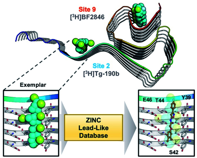
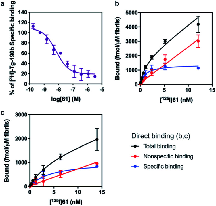
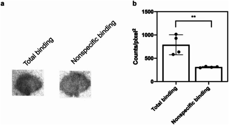
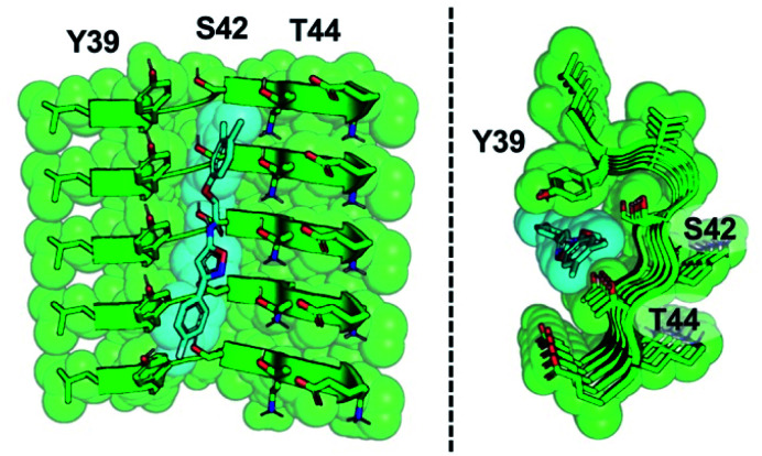
Small molecules that bind with high affinity and specificity to fibrils of the α-synuclein (αS) protein have the potential to serve as positron emission tomography (PET) imaging probes to aid in the diagnosis of Parkinson's disease and related synucleinopathies. To identify such molecules, we employed an ultra-high throughput screening strategy using idealized pseudo-ligands termed exemplars to identify compounds for experimental binding studies. For the top hit from this screen, we used photo-crosslinking to confirm its binding site and studied the structure-activity relationship of its analogs to develop multiple molecules with nanomolar affinity for αS fibrils and moderate specificity for αS over Aβ fibrils. Lastly, we demonstrated the potential of the lead analog as an imaging probe by measuring binding to αS-enriched homogenates from mouse brain tissue using a radiolabeled analog of the identified molecule. This study demonstrates the validity of our powerful new approach to the discovery of PET probes for challenging molecular targets.

摘要

与α-突触核蛋白(αS)纤维具有高亲和力和特异性结合的小分子,有潜力作为正电子发射断层扫描(PET)成像探针,辅助帕金森病及相关突触核蛋白病的诊断。为了鉴定此类分子,我们采用了一种超高通量筛选策略,使用称为范例的理想化假配体来鉴定用于实验性结合研究的化合物。对于此次筛选中最突出的结果,我们利用光交联来确认其结合位点,并研究其类似物的构效关系,以开发出多种对αS纤维具有纳摩尔亲和力且对αS比对Aβ纤维具有适度特异性的分子。最后,我们通过使用所鉴定分子的放射性标记类似物测量其与来自小鼠脑组织的富含αS的匀浆的结合,证明了先导类似物作为成像探针的潜力。这项研究证明了我们强大的新方法对于发现针对具有挑战性的分子靶点的PET探针的有效性。

https://cdn.ncbi.nlm.nih.gov/pmc/blobs/9745/8047729/ff0c09ac9993/d0sc02159h-f7.jpg
https://cdn.ncbi.nlm.nih.gov/pmc/blobs/9745/8047729/a50a8e8264c7/d0sc02159h-f1.jpg
https://cdn.ncbi.nlm.nih.gov/pmc/blobs/9745/8047729/0b26540545d2/d0sc02159h-f2.jpg
https://cdn.ncbi.nlm.nih.gov/pmc/blobs/9745/8047729/df0b506bb03b/d0sc02159h-f3.jpg
https://cdn.ncbi.nlm.nih.gov/pmc/blobs/9745/8047729/f9544094f5a2/d0sc02159h-s1.jpg
https://cdn.ncbi.nlm.nih.gov/pmc/blobs/9745/8047729/94b6a0940fac/d0sc02159h-f4.jpg
https://cdn.ncbi.nlm.nih.gov/pmc/blobs/9745/8047729/2dee9df4798e/d0sc02159h-f5.jpg
https://cdn.ncbi.nlm.nih.gov/pmc/blobs/9745/8047729/35c12166020f/d0sc02159h-f6.jpg
https://cdn.ncbi.nlm.nih.gov/pmc/blobs/9745/8047729/ff0c09ac9993/d0sc02159h-f7.jpg

相似文献

1
Identification of a nanomolar affinity α-synuclein fibril imaging probe by ultra-high throughput screening.通过超高通量筛选鉴定纳摩尔亲和力的α-突触核蛋白原纤维成像探针。
Chem Sci. 2020 Sep 10;11(47):12746-12754. doi: 10.1039/d0sc02159h. eCollection 2020 Dec 21.
2
Identification of a Putative α-synuclein Radioligand Using an in silico Similarity Search.利用计算机模拟相似性搜索鉴定潜在的α-突触核蛋白放射性配体。
Mol Imaging Biol. 2023 Aug;25(4):704-719. doi: 10.1007/s11307-023-01814-9. Epub 2023 Mar 29.
3
A KLVFFAE-Derived Peptide Probe for Detection of Alpha-Synuclein Fibrils.一种用于检测α-突触核蛋白纤维的 KLVFFAE 衍生肽探针。
Appl Biochem Biotechnol. 2020 Apr;190(4):1411-1424. doi: 10.1007/s12010-019-03197-6. Epub 2019 Nov 27.
4
SARS-CoV-2 N-protein induces the formation of composite α-synuclein/N-protein fibrils that transform into a strain of α-synuclein fibrils.SARS-CoV-2 N 蛋白诱导形成复合α-突触核蛋白/N 蛋白纤维,这些纤维转化为一种α-突触核蛋白纤维。
Nanoscale. 2023 Nov 23;15(45):18337-18346. doi: 10.1039/d3nr03556e.
5
NMR unveils an N-terminal interaction interface on acetylated-α-synuclein monomers for recruitment to fibrils.NMR 揭示乙酰化-α-突触核蛋白单体上的 N 端相互作用界面,用于招募到纤维中。
Proc Natl Acad Sci U S A. 2021 May 4;118(18). doi: 10.1073/pnas.2017452118.
6
The effect of truncation on prion-like properties of α-synuclein.截断对α-突触核蛋白类朊病毒特性的影响。
J Biol Chem. 2018 Sep 7;293(36):13910-13920. doi: 10.1074/jbc.RA118.001862. Epub 2018 Jul 20.
7
Phenolic compounds prevent the oligomerization of α-synuclein and reduce synaptic toxicity.酚类化合物可防止α-突触核蛋白寡聚化并降低突触毒性。
J Neurochem. 2015 Sep;134(5):943-55. doi: 10.1111/jnc.13180. Epub 2015 Jun 17.
8
In Silico, in Vitro, and in Vivo Evaluation of New Candidates for α-Synuclein PET Imaging.新型α-突触核蛋白 PET 成像示踪剂的计算机模拟、体外和体内评价。
Mol Pharm. 2018 Aug 6;15(8):3153-3166. doi: 10.1021/acs.molpharmaceut.8b00229. Epub 2018 Jul 25.
9
Comparison of the in vivo induction and transmission of α-synuclein pathology by mutant α-synuclein fibril seeds in transgenic mice.比较突变α-突触核蛋白纤维种子在转基因小鼠体内诱导和传播α-突触核蛋白病理的情况。
Hum Mol Genet. 2017 Dec 15;26(24):4906-4915. doi: 10.1093/hmg/ddx371.
10
Increased Dynamics of α-Synuclein Fibrils by β-Synuclein Leads to Reduced Seeding and Cytotoxicity.β-突触核蛋白导致α-突触核蛋白纤维动态增加,从而降低了种子形成和细胞毒性。
Sci Rep. 2019 Nov 26;9(1):17579. doi: 10.1038/s41598-019-54063-8.

引用本文的文献

1
Genetic code expansion and enzymatic modifications as accessible methods for studying site-specific post-translational modifications of alpha-synuclein and tau.遗传密码扩展和酶促修饰作为研究α-突触核蛋白和tau蛋白位点特异性翻译后修饰的可行方法。
Protein Sci. 2025 Oct;34(10):e70302. doi: 10.1002/pro.70302.
2
Alpha-Synuclein Fibril Structures Cluster into Distinct Classes.α-突触核蛋白原纤维结构聚集成不同类别。
bioRxiv. 2025 May 2:2025.04.30.651534. doi: 10.1101/2025.04.30.651534.
3
Detecting alpha-synuclein aggregates with small molecules on single-molecule array.

本文引用的文献

1
Structures of α-synuclein filaments from multiple system atrophy.多系统萎缩中α-突触核蛋白丝的结构。
Nature. 2020 Sep;585(7825):464-469. doi: 10.1038/s41586-020-2317-6. Epub 2020 May 27.
2
Structural heterogeneity of α-synuclein fibrils amplified from patient brain extracts.从患者脑组织提取物中放大的α-突触核蛋白纤维的结构异质性。
Nat Commun. 2019 Dec 4;10(1):5535. doi: 10.1038/s41467-019-13564-w.
3
Functional cooperation of α-synuclein and VAMP2 in synaptic vesicle recycling.α-突触核蛋白与 VAMP2 在突触囊泡循环中的功能合作。
在单分子阵列上用小分子检测α-突触核蛋白聚集体。
Chem Sci. 2025 Jun 16. doi: 10.1039/d4sc07649d.
4
Development of Positron Emission Tomography Radiotracers for Imaging α-Synuclein Aggregates.用于α-突触核蛋白聚集体成像的正电子发射断层显像剂的研发
Cells. 2025 Jun 16;14(12):907. doi: 10.3390/cells14120907.
5
Ligands for Protein Fibrils of Amyloid-β, α-Synuclein, and Tau.淀粉样β蛋白、α-突触核蛋白和tau蛋白原纤维的配体
Chem Rev. 2025 Jun 11;125(11):5282-5348. doi: 10.1021/acs.chemrev.4c00838. Epub 2025 May 6.
6
The development of a PET radiotracer for imaging alpha synuclein aggregates in Parkinson's disease.用于帕金森病中α-突触核蛋白聚集体成像的正电子发射断层显像(PET)放射性示踪剂的研发。
RSC Med Chem. 2025 Apr 3. doi: 10.1039/d5md00057b.
7
Strategic Modulation of Polarity and Viscosity Sensitivity of Bimane Molecular Rotor-Based Fluorophores for Imaging α-Synuclein.用于成像α-突触核蛋白的基于双萘嵌苯分子转子的荧光团的极性和粘度敏感性的策略性调控
bioRxiv. 2025 Jan 8:2025.01.07.631748. doi: 10.1101/2025.01.07.631748.
8
Strategies of positron emission tomography (PET) tracer development for imaging of tau and α-synuclein in neurodegenerative disorders.用于神经退行性疾病中tau蛋白和α-突触核蛋白成像的正电子发射断层扫描(PET)示踪剂开发策略。
RSC Med Chem. 2024 Nov 20. doi: 10.1039/d4md00576g.
9
Binding adaptability of chemical ligands to polymorphic α-synuclein amyloid fibrils.化学配体与多态α-突触核蛋白淀粉样纤维的结合适应性。
Proc Natl Acad Sci U S A. 2024 Aug 27;121(35):e2321633121. doi: 10.1073/pnas.2321633121. Epub 2024 Aug 22.
10
Efficient characterization of multiple binding sites of small molecule imaging ligands on amyloid-beta, tau and alpha-synuclein.高效鉴定小分子成像配体在淀粉样蛋白-β、tau 和 α-突触核蛋白上的多个结合位点。
Eur J Nucl Med Mol Imaging. 2024 Nov;51(13):3960-3977. doi: 10.1007/s00259-024-06806-7. Epub 2024 Jul 2.
Proc Natl Acad Sci U S A. 2019 Jun 4;116(23):11113-11115. doi: 10.1073/pnas.1903049116. Epub 2019 May 20.
4
Synapsins regulate α-synuclein functions.突触结合蛋白调节α-突触核蛋白的功能。
Proc Natl Acad Sci U S A. 2019 Jun 4;116(23):11116-11118. doi: 10.1073/pnas.1903054116. Epub 2019 May 20.
5
Chalcones and Five-Membered Heterocyclic Isosteres Bind to Alpha Synuclein Fibrils in Vitro.查尔酮和五元杂环电子等排体在体外与α-突触核蛋白原纤维结合。
ACS Omega. 2018 Apr 30;3(4):4486-4493. doi: 10.1021/acsomega.7b01897. Epub 2018 Apr 24.
6
Cryo-EM of full-length α-synuclein reveals fibril polymorphs with a common structural kernel.全长α-突触核蛋白的冷冻电镜解析揭示了具有共同结构核心的纤维多态性。
Nat Commun. 2018 Sep 6;9(1):3609. doi: 10.1038/s41467-018-05971-2.
7
Structures of filaments from Pick's disease reveal a novel tau protein fold.Pick 病纤维结构揭示了一种新型的 tau 蛋白折叠。
Nature. 2018 Sep;561(7721):137-140. doi: 10.1038/s41586-018-0454-y. Epub 2018 Aug 29.
8
Amyloid fibril structure of α-synuclein determined by cryo-electron microscopy.通过冷冻电子显微镜确定α-突触核蛋白的淀粉样纤维结构。
Cell Res. 2018 Sep;28(9):897-903. doi: 10.1038/s41422-018-0075-x. Epub 2018 Jul 31.
9
Cryo-EM structure of alpha-synuclein fibrils.α-突触核蛋白纤维的冷冻电镜结构。
Elife. 2018 Jul 3;7:e36402. doi: 10.7554/eLife.36402.
10
A Protofilament-Protofilament Interface in the Structure of Mouse α-Synuclein Fibrils.鼠源α-突触核蛋白纤维结构中的原纤维-原纤维界面。
Biophys J. 2018 Jun 19;114(12):2811-2819. doi: 10.1016/j.bpj.2018.05.011.