• 文献检索
  • 文档翻译
  • 深度研究
  • 学术资讯
  • Suppr Zotero 插件Zotero 插件
  • 邀请有礼
  • 套餐&价格
  • 历史记录
应用&插件
Suppr Zotero 插件Zotero 插件浏览器插件Mac 客户端Windows 客户端微信小程序
定价
高级版会员购买积分包购买API积分包
服务
文献检索文档翻译深度研究API 文档MCP 服务
关于我们
关于 Suppr公司介绍联系我们用户协议隐私条款
关注我们

Suppr 超能文献

核心技术专利:CN118964589B侵权必究
粤ICP备2023148730 号-1Suppr @ 2026

文献检索

告别复杂PubMed语法,用中文像聊天一样搜索,搜遍4000万医学文献。AI智能推荐,让科研检索更轻松。

立即免费搜索

文件翻译

保留排版,准确专业,支持PDF/Word/PPT等文件格式,支持 12+语言互译。

免费翻译文档

深度研究

AI帮你快速写综述,25分钟生成高质量综述,智能提取关键信息,辅助科研写作。

立即免费体验

黏附连接的参与调节心脏起搏器细胞谱系的功能模式。

Adherens junction engagement regulates functional patterning of the cardiac pacemaker cell lineage.

作者信息

Thomas Kandace, Henley Trevor, Rossi Simone, Costello M Joseph, Polacheck William, Griffith Boyce E, Bressan Michael

机构信息

Department of Cell Biology and Physiology, University of North Carolina at Chapel Hill, Chapel Hill, NC 27599, USA; McAllister Heart Institute, University of North Carolina at Chapel Hill, Chapel Hill, NC 27599, USA.

Department of Mathematics, University of North Carolina, Chapel Hill, NC, USA.

出版信息

Dev Cell. 2021 May 17;56(10):1498-1511.e7. doi: 10.1016/j.devcel.2021.04.004. Epub 2021 Apr 22.

DOI:10.1016/j.devcel.2021.04.004
PMID:33891897
原文链接:https://pmc.ncbi.nlm.nih.gov/articles/PMC8137639/
Abstract

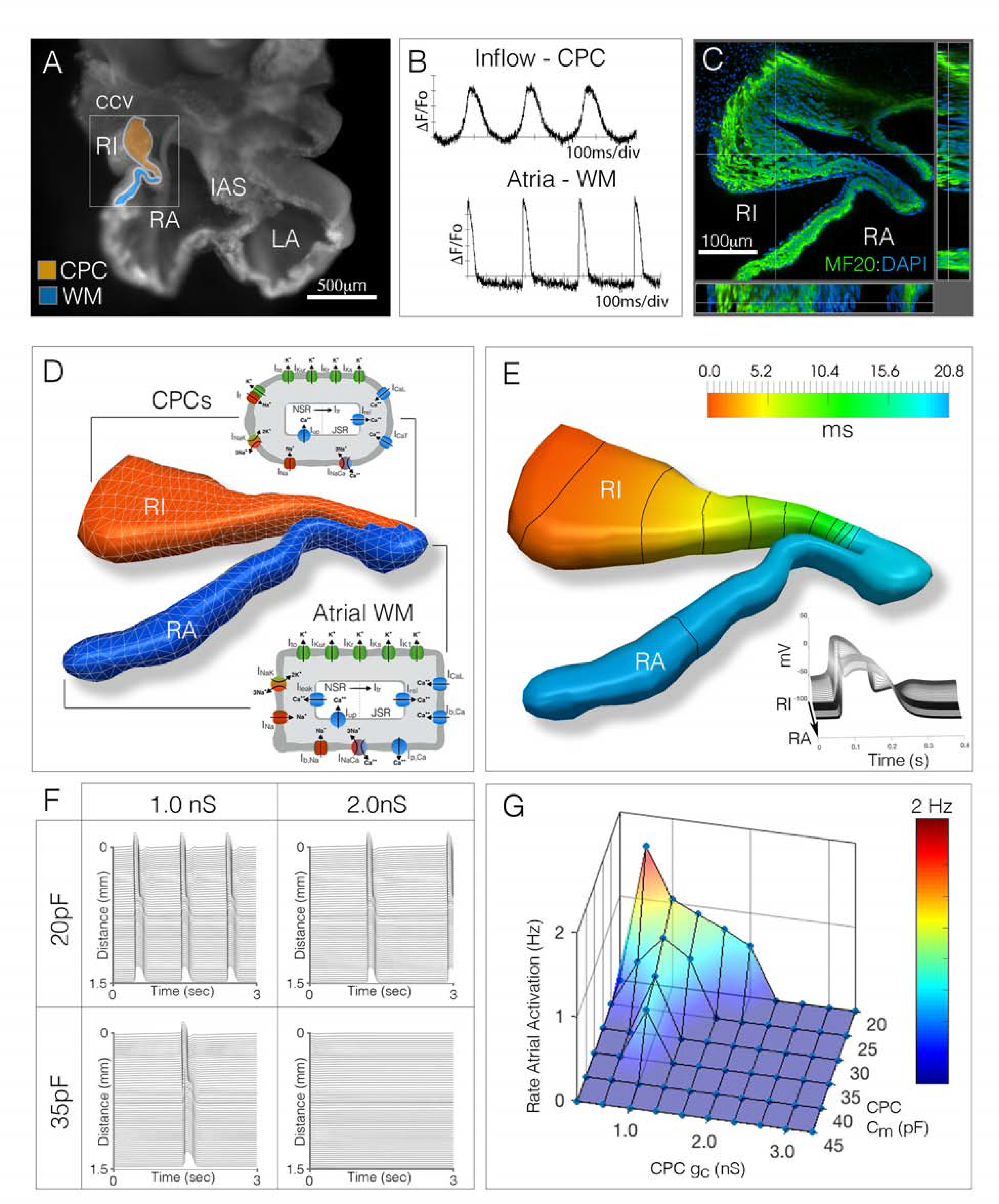
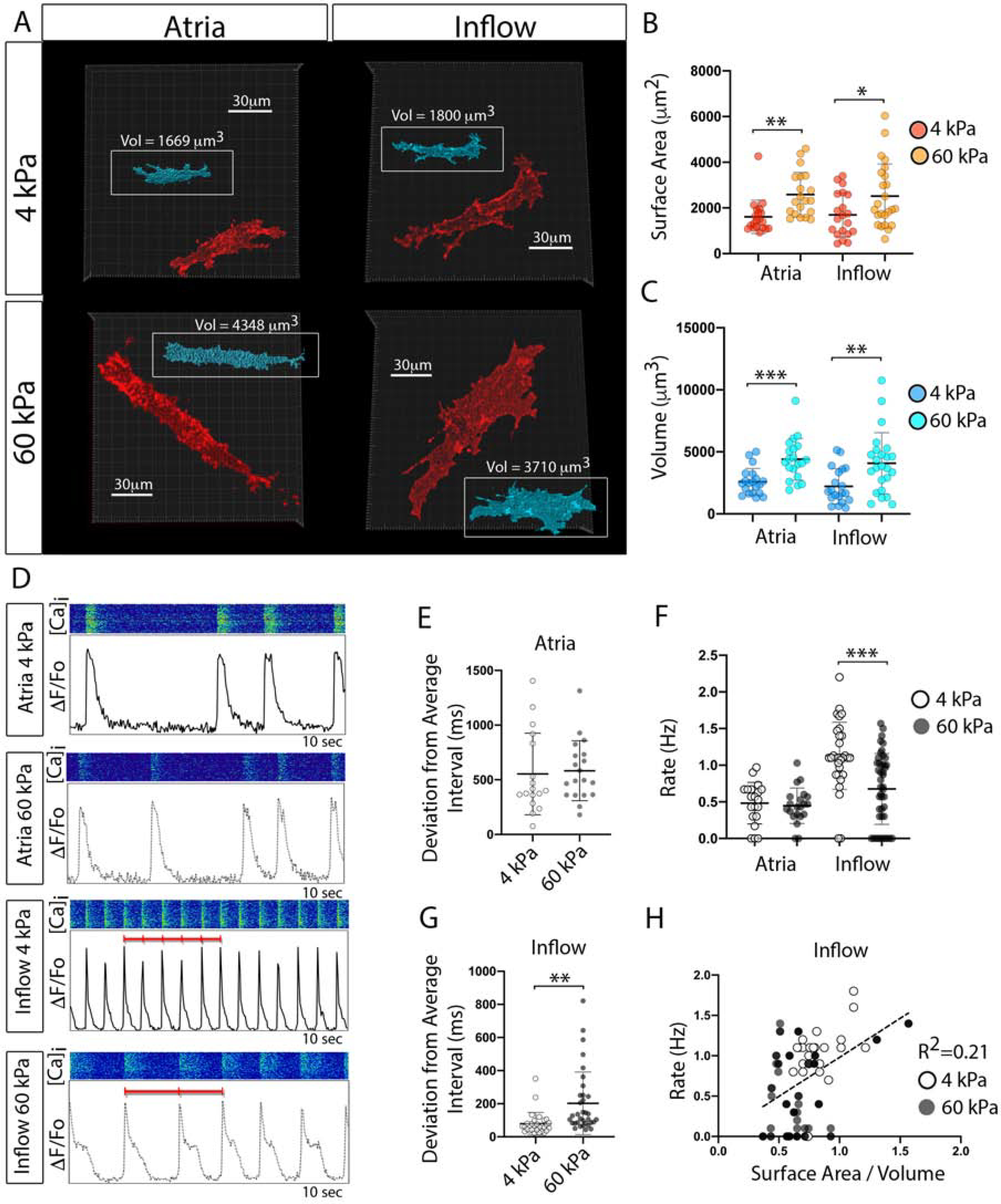
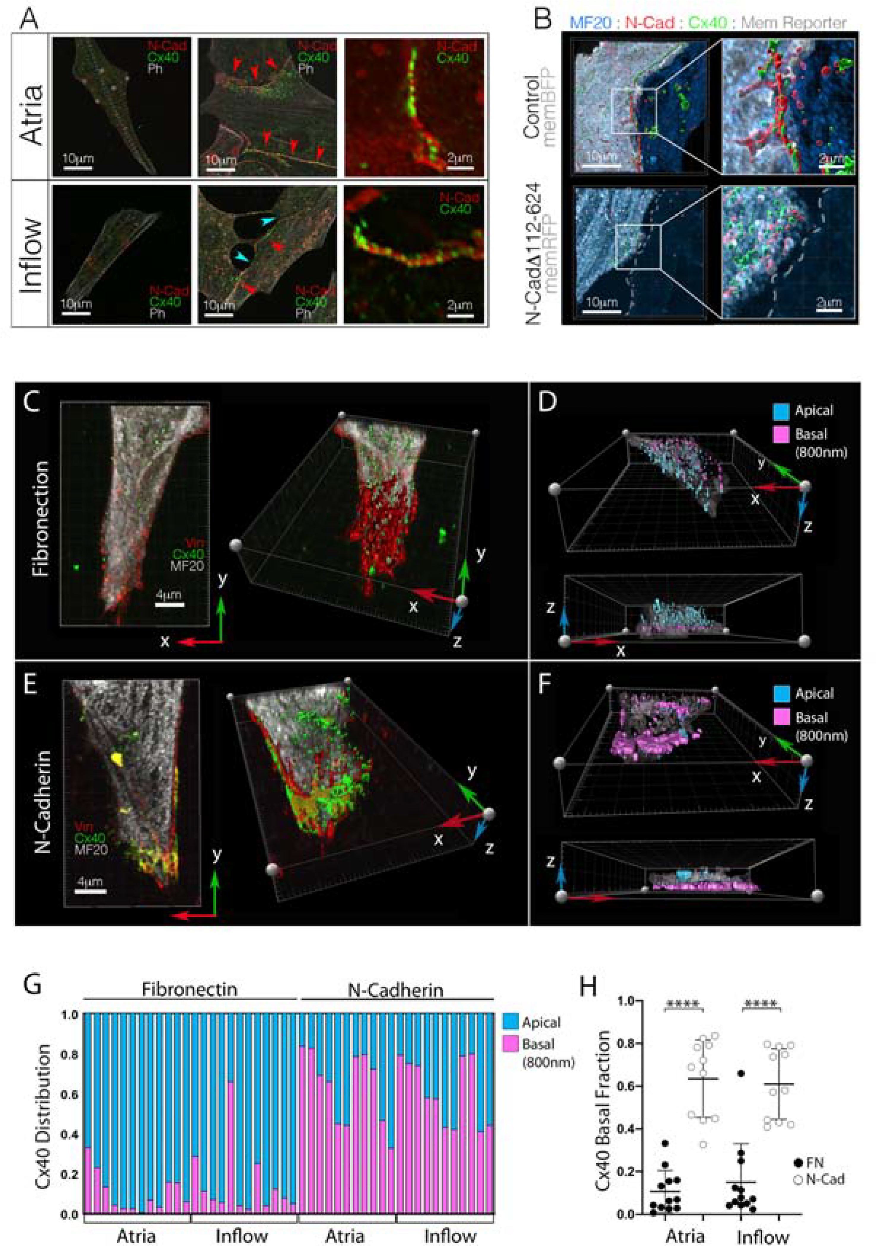
Cardiac pacemaker cells (CPCs) rhythmically initiate the electrical impulses that drive heart contraction. CPCs display the highest rate of spontaneous depolarization in the heart despite being subjected to inhibitory electrochemical conditions that should theoretically suppress their activity. While several models have been proposed to explain this apparent paradox, the actual molecular mechanisms that allow CPCs to overcome electrogenic barriers to their function remain poorly understood. Here, we have traced CPC development at single-cell resolution and uncovered a series of cytoarchitectural patterning events that are critical for proper pacemaking. Specifically, our data reveal that CPCs dynamically modulate adherens junction (AJ) engagement to control characteristics including surface area, volume, and gap junctional coupling. This allows CPCs to adopt a structural configuration that supports their overall excitability. Thus, our data have identified a direct role for local cellular mechanics in patterning critical morphological features that are necessary for CPC electrical activity.

摘要

心脏起搏器细胞(CPCs)有节奏地引发驱动心脏收缩的电冲动。尽管处于理论上应抑制其活动的抑制性电化学条件下,但CPCs在心脏中表现出最高的自发去极化速率。虽然已经提出了几种模型来解释这一明显的矛盾现象,但对于使CPCs克服其功能的电生障碍的实际分子机制仍知之甚少。在这里,我们以单细胞分辨率追踪了CPCs的发育过程,并发现了一系列对于正常起搏至关重要的细胞结构模式形成事件。具体而言,我们的数据显示,CPCs动态调节黏附连接(AJ)的参与,以控制包括表面积、体积和间隙连接耦合等特征。这使得CPCs能够采用一种支持其整体兴奋性的结构构型。因此,我们的数据确定了局部细胞力学在塑造CPCs电活动所需的关键形态特征方面的直接作用。

https://cdn.ncbi.nlm.nih.gov/pmc/blobs/871f/8137639/0a998b71dcc7/nihms-1694322-f0008.jpg
https://cdn.ncbi.nlm.nih.gov/pmc/blobs/871f/8137639/da63c2cbdc49/nihms-1694322-f0002.jpg
https://cdn.ncbi.nlm.nih.gov/pmc/blobs/871f/8137639/e92baa843e05/nihms-1694322-f0003.jpg
https://cdn.ncbi.nlm.nih.gov/pmc/blobs/871f/8137639/ee8d2c793205/nihms-1694322-f0004.jpg
https://cdn.ncbi.nlm.nih.gov/pmc/blobs/871f/8137639/0ee7d5037647/nihms-1694322-f0005.jpg
https://cdn.ncbi.nlm.nih.gov/pmc/blobs/871f/8137639/91ffbf920938/nihms-1694322-f0006.jpg
https://cdn.ncbi.nlm.nih.gov/pmc/blobs/871f/8137639/4d4eb12c89ed/nihms-1694322-f0007.jpg
https://cdn.ncbi.nlm.nih.gov/pmc/blobs/871f/8137639/0a998b71dcc7/nihms-1694322-f0008.jpg

相似文献

1
Adherens junction engagement regulates functional patterning of the cardiac pacemaker cell lineage.黏附连接的参与调节心脏起搏器细胞谱系的功能模式。
Dev Cell. 2021 May 17;56(10):1498-1511.e7. doi: 10.1016/j.devcel.2021.04.004. Epub 2021 Apr 22.
2
Local tissue mechanics control cardiac pacemaker cell embryonic patterning.局部组织力学控制心脏起搏细胞胚胎模式形成。
Life Sci Alliance. 2023 Mar 27;6(6). doi: 10.26508/lsa.202201799. Print 2023 Jun.
3
Structural and Intermolecular Associations Between Connexin36 and Protein Components of the Adherens Junction-Neuronal Gap Junction Complex.连接蛋白 36 与黏着连接-神经元缝隙连接复合体质子成分之间的结构和分子间的联系。
Neuroscience. 2018 Aug 1;384:241-261. doi: 10.1016/j.neuroscience.2018.05.026. Epub 2018 Jun 5.
4
The organization of adherens junctions and desmosomes at the cardiac intercalated disc is independent of gap junctions.心脏闰盘处黏着连接和桥粒的组织与间隙连接无关。
J Cell Sci. 2003 Mar 1;116(Pt 5):875-85. doi: 10.1242/jcs.00258.
5
Assembly of the cardiac intercalated disk during pre- and postnatal development of the human heart.人类心脏产前和产后发育过程中心脏闰盘的组装。
PLoS One. 2014 Apr 14;9(4):e94722. doi: 10.1371/journal.pone.0094722. eCollection 2014.
6
Intercalated discs: cellular adhesion and signaling in heart health and diseases.连接蛋白:心脏健康与疾病中的细胞黏附和信号转导。
Heart Fail Rev. 2019 Jan;24(1):115-132. doi: 10.1007/s10741-018-9743-7.
7
Loss of αT-catenin alters the hybrid adhering junctions in the heart and leads to dilated cardiomyopathy and ventricular arrhythmia following acute ischemia.αT-连环蛋白的缺失改变了心脏中的混合黏附连接,导致急性缺血后扩张型心肌病和室性心律失常。
J Cell Sci. 2012 Feb 15;125(Pt 4):1058-67. doi: 10.1242/jcs.098640. Epub 2012 Mar 15.
8
Reduced expression of adherens and gap junction proteins can have a fundamental role in the development of heart failure following cardiac hypertrophy in rats.在大鼠心脏肥大后发生心力衰竭的过程中,黏附连接蛋白和缝隙连接蛋白表达降低可能起到根本性作用。
Exp Mol Pathol. 2016 Feb;100(1):167-76. doi: 10.1016/j.yexmp.2015.12.009. Epub 2015 Dec 17.
9
Dissociated spatial patterning of gap junctions and cell adhesion junctions during postnatal differentiation of ventricular myocardium.出生后心室心肌分化过程中缝隙连接和细胞黏附连接的解离空间模式
Circ Res. 1997 Jan;80(1):88-94. doi: 10.1161/01.res.80.1.88.
10
A common Shox2-Nkx2-5 antagonistic mechanism primes the pacemaker cell fate in the pulmonary vein myocardium and sinoatrial node.一种常见的Shox2-Nkx2-5拮抗机制决定了肺静脉心肌和窦房结中起搏细胞的命运。
Development. 2015 Jul 15;142(14):2521-32. doi: 10.1242/dev.120220. Epub 2015 Jul 2.

引用本文的文献

1
The components of an electrical synapse as revealed by expansion microscopy of a single synaptic contact.通过对单个突触接触的扩展显微镜观察揭示电突触的组成部分。
Elife. 2024 Jul 12;13:e91931. doi: 10.7554/eLife.91931.
2
Biomimetic Cardiac Tissue Models for In Vitro Arrhythmia Studies.用于体外心律失常研究的仿生心脏组织模型
Biomimetics (Basel). 2023 Oct 14;8(6):487. doi: 10.3390/biomimetics8060487.
3
Local tissue mechanics control cardiac pacemaker cell embryonic patterning.局部组织力学控制心脏起搏细胞胚胎模式形成。

本文引用的文献

1
Desmin is essential for the structure and function of the sinoatrial node: implications for increased arrhythmogenesis.结蛋白对于窦房结的结构和功能至关重要:增加心律失常发生的影响。
Am J Physiol Heart Circ Physiol. 2020 Sep 1;319(3):H557-H570. doi: 10.1152/ajpheart.00594.2019. Epub 2020 Jul 17.
2
Engineered Cardiac Pacemaker Nodes Created by TBX18 Gene Transfer Overcome Source-Sink Mismatch.通过TBX18基因转移构建的工程化心脏起搏器节点克服了源-汇不匹配问题。
Adv Sci (Weinh). 2019 Sep 30;6(22):1901099. doi: 10.1002/advs.201901099. eCollection 2019 Nov.
3
Vinculin anchors contractile actin to the cardiomyocyte adherens junction.
Life Sci Alliance. 2023 Mar 27;6(6). doi: 10.26508/lsa.202201799. Print 2023 Jun.
4
Perinatal Lead Exposure Promotes Sex-Specific Epigenetic Programming of Disease-Relevant Pathways in Mouse Heart.围产期铅暴露促进小鼠心脏疾病相关通路的性别特异性表观遗传编程。
Toxics. 2023 Jan 16;11(1):85. doi: 10.3390/toxics11010085.
5
Novel Insights into the Sinoatrial Node in Single-Cell RNA Sequencing: From Developmental Biology to Physiological Function.单细胞RNA测序中对窦房结的新见解:从发育生物学到生理功能
J Cardiovasc Dev Dis. 2022 Nov 18;9(11):402. doi: 10.3390/jcdd9110402.
6
Transcriptional and Epigenetic Landscape of Cardiac Pacemaker Cells: Insights Into Cellular Specialization in the Sinoatrial Node.心脏起搏器细胞的转录和表观遗传图谱:对窦房结细胞特化的见解
Front Physiol. 2021 Jul 16;12:712666. doi: 10.3389/fphys.2021.712666. eCollection 2021.
粘着斑蛋白将收缩性肌动蛋白锚定到心肌细胞黏着连接。
Mol Biol Cell. 2019 Oct 1;30(21):2639-2650. doi: 10.1091/mbc.E19-04-0216. Epub 2019 Sep 4.
4
Simplified platform for mosaic in vivo analysis of cellular maturation in the developing heart.简化的体内镶嵌分析平台,用于研究心脏发育过程中的细胞成熟。
Sci Rep. 2019 Jul 24;9(1):10716. doi: 10.1038/s41598-019-47009-7.
5
Quantitative proteomics and single-nucleus transcriptomics of the sinus node elucidates the foundation of cardiac pacemaking.窦房结的定量蛋白质组学和单核转录组学阐明了心脏起搏的基础。
Nat Commun. 2019 Jun 28;10(1):2889. doi: 10.1038/s41467-019-10709-9.
6
Transcriptome analysis of mouse and human sinoatrial node cells reveals a conserved genetic program.对小鼠和人类窦房结细胞的转录组分析揭示了一个保守的遗传程序。
Development. 2019 Apr 25;146(8):dev173161. doi: 10.1242/dev.173161.
7
Dynamic Cellular Integration Drives Functional Assembly of the Heart's Pacemaker Complex.动态细胞整合驱动心脏起搏器复合体的功能组装。
Cell Rep. 2018 May 22;23(8):2283-2291. doi: 10.1016/j.celrep.2018.04.075.
8
α-Catenin-dependent cytoskeletal tension controls Yap activity in the heart.α-连环蛋白依赖性细胞骨架张力控制心脏中的Yap活性。
Development. 2018 Mar 8;145(5):dev149823. doi: 10.1242/dev.149823.
9
Incorporating inductances in tissue-scale models of cardiac electrophysiology.将电感纳入心脏电生理学的组织尺度模型中。
Chaos. 2017 Sep;27(9):093926. doi: 10.1063/1.5000706.
10
Electrophysiological heterogeneity of pacemaker cells in the rabbit intercaval region, including the SA node: insights from recording multiple ion currents in each cell.兔腔静脉区起搏细胞(包括窦房结)的电生理异质性:在每个细胞中记录多种离子流的研究结果
Am J Physiol Heart Circ Physiol. 2018 Mar 1;314(3):H403-H414. doi: 10.1152/ajpheart.00253.2016. Epub 2017 Sep 15.