Department of Radiodiagnosis, All India Institute of Medical Sciences, Patna, Bihar, India.
Department of Gastroenterology, All India Institute of Medical Sciences, Patna, Bihar, India.
Abdom Radiol (NY). 2021 Sep;46(9):4148-4158. doi: 10.1007/s00261-021-03093-w. Epub 2021 Apr 24.
To identify different morphological types of amebic liver abscess (ALA) based on CT findings and to assess whether they have different clinical features.
CT images of 112 symptomatic patients with ALA were analyzed to identify the imaging features distinctive of each morphological type. The following CT findings were investigated: the presence of abscess wall, rim enhancement, edge characteristic, septa, intermediate density zone, and peripheral hypodensity. Abscesses from each type were further evaluated for their clinical presentations, laboratory findings and outcomes.
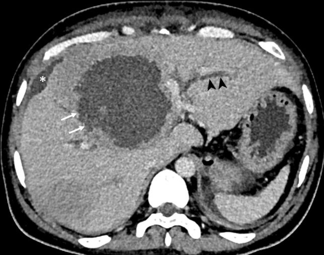
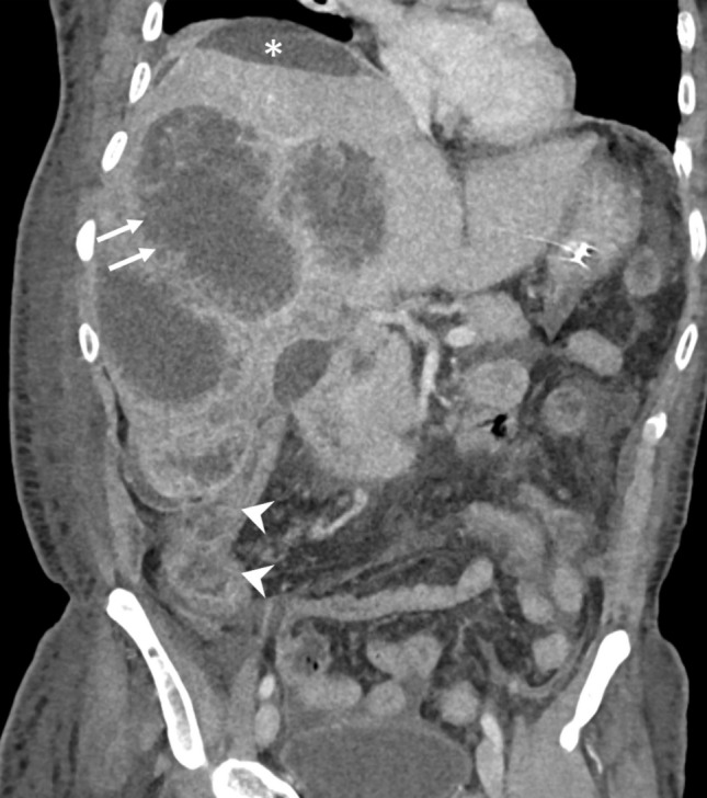
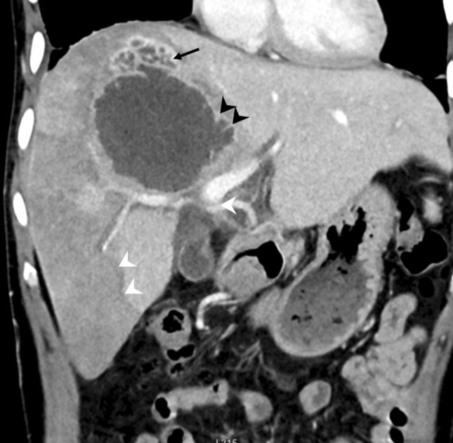
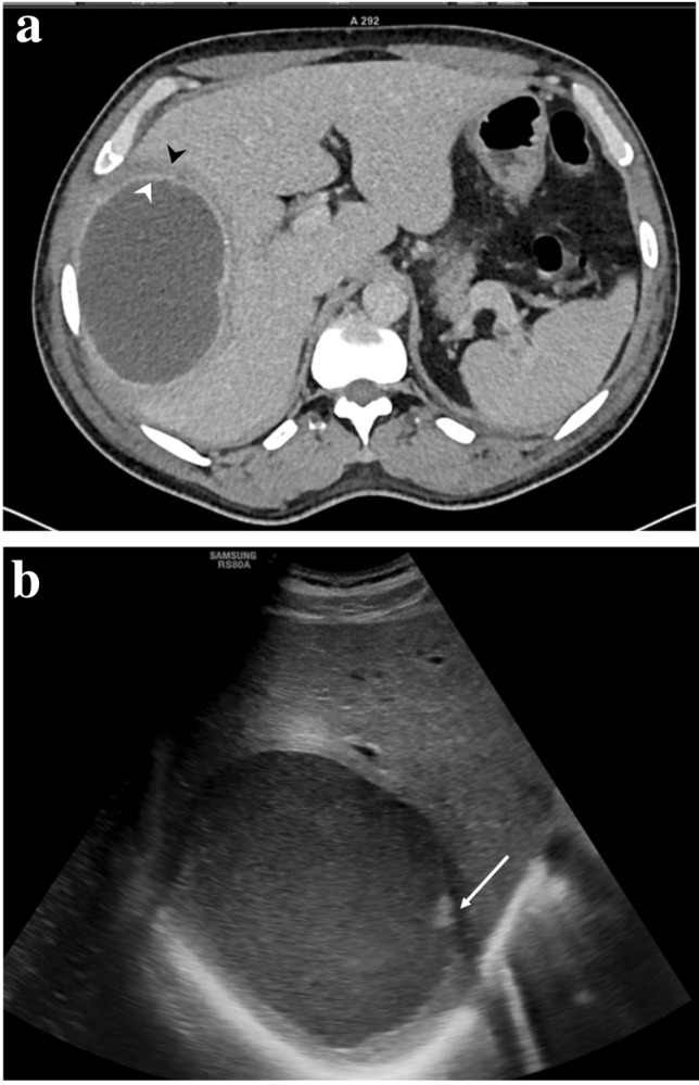
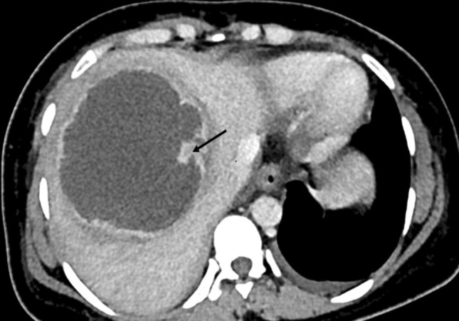
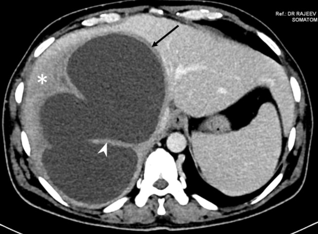
We identified three types of ALAs: type I, II and III. Type I abscesses (66%) were characterized by absent or incomplete walls, ragged edges and peripheral septa; their edges exhibited irregular and interrupted enhancement. Type II (28%) had a complete wall characterized by rim enhancement and peripheral hypodense halo. Type III (6%) demonstrated a wall but without enhancement. Clinically, type I abscesses presented acutely with severe disease. They had significantly deranged laboratory parameters, higher incidence of rupture and higher rate of inpatient or intensive care unit admission. The severity of the disease prompted immediate percutaneous drainage in most type I abscesses (81%). Two of them died from multiple organ failure. The type II or III abscesses, on the other hand, had delayed presentations with mild to moderate disease, with near normal laboratory findings.
ALAs have three different CT morphological types, with different clinical and laboratory features. Percutaneous drainage is indicated in most of type I abscesses.
根据 CT 表现识别阿米巴肝脓肿(ALA)的不同形态类型,并评估它们是否具有不同的临床特征。
分析 112 例有症状的 ALA 患者的 CT 图像,以识别每种形态类型的特征性影像学表现。研究的 CT 表现包括:脓肿壁的存在、边缘强化、边缘特征、分隔、中间密度区和外周低密度区。对每种类型的脓肿进一步评估其临床表现、实验室检查结果和转归。
我们确定了三种类型的 ALA:I 型、II 型和 III 型。I 型脓肿(66%)表现为无壁或不完全壁、边缘不整和外周分隔;其边缘呈不规则和间断性强化。II 型(28%)具有完整的壁,表现为边缘强化和外周低密度晕环。III 型(6%)表现为有壁但无强化。临床上,I 型脓肿表现为急性起病,病情严重。实验室参数明显异常,破裂发生率高,住院或入住重症监护病房的比例高。由于疾病严重,大多数 I 型脓肿(81%)立即行经皮引流。其中 2 例死于多器官功能衰竭。另一方面,II 型或 III 型脓肿表现为起病较晚,病情轻至中度,实验室检查结果接近正常。
ALA 有三种不同的 CT 形态类型,具有不同的临床和实验室特征。大多数 I 型脓肿需要经皮引流。