Department of Neuroscience, Neurosurgery, Uppsala University, Uppsala, Sweden; Lund University, Skåne University Hospital, Department of Clinical Sciences Lund, Neurosurgery, Lund, Sweden.
Department of Neuroscience, Neurosurgery, Uppsala University, Uppsala, Sweden.
Neuroimage Clin. 2021;30:102665. doi: 10.1016/j.nicl.2021.102665. Epub 2021 Apr 7.
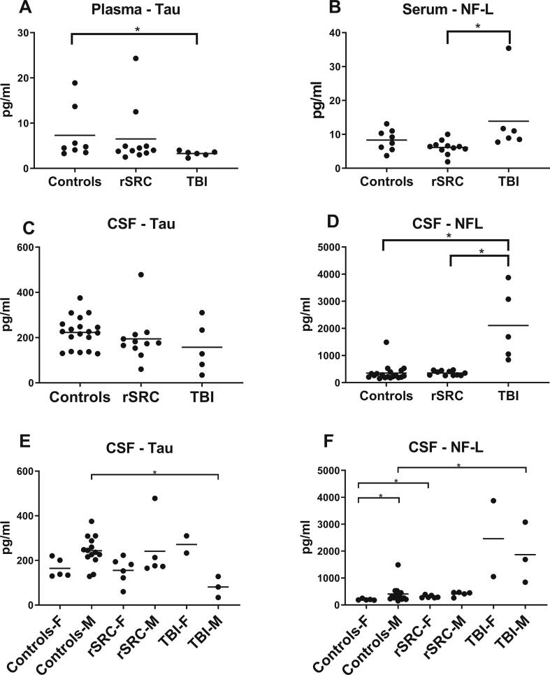
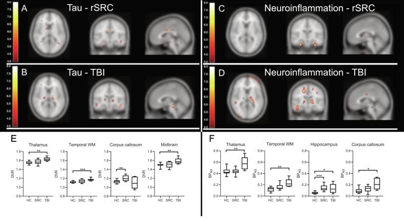
Traumatic brain injury (TBI) and repeated sports-related concussions (rSRCs) are associated with an increased risk for neurodegeneration. Autopsy findings of selected cohorts of long-term TBI survivors and rSRC athletes reveal increased tau aggregation and a persistent neuroinflammation. To assess in vivo tau aggregation and neuroinflammation in young adult TBI and rSRC cohorts, we evaluated 9 healthy controls (mean age 26 ± 5 years; 4 males, 5 females), 12 symptomatic athletes (26 ± 7 years; 6 males, 6 females) attaining ≥3 previous SRCs, and 6 moderate-to severe TBI patients (27 ± 7 years; 4 males, 2 females) in a combined positron emission tomography (PET)/magnetic resonance (MR) scanner ≥6 months post-injury. Dual PET tracers, [F]THK5317 for tau aggregation and [C]PK11195 for neuroinflammation/microglial activation, were investigated on the same day. The Repeated Battery Assessment of Neurological Status (RBANS) scores, used for cognitive evaluation, were lower in both the rSRC and TBI groups (p < 0.05). Neurofilament-light (NF-L) levels were increased in plasma and cerebrospinal fluid (CSF; p < 0.05), and serum tau levels lower, in TBI although not in rSRC. In rSRC athletes, PET imaging showed increased neuroinflammation in the hippocampus and tau aggregation in the corpus callosum. In TBI patients, tau aggregation was observed in thalami, temporal white matter and midbrain; widespread neuroinflammation was found e.g. in temporal white matter, hippocampus and corpus callosum. In mixed-sex cohorts of young adult athletes with persistent post-concussion symptoms and in TBI patients, increased tau aggregation and neuroinflammation are observed at ≥6 months post-injury using PET. Studies with extended clinical follow-up, biomarker examinations and renewed PET imaging are needed to evaluate whether these findings progress to a neurodegenerative disorder or if spontaneous resolution is possible.
创伤性脑损伤(TBI)和反复的与运动相关的脑震荡(rSRC)与神经退行性变的风险增加有关。对长期 TBI 幸存者和 rSRC 运动员的选定队列的尸检结果显示,tau 聚集增加和持续的神经炎症。为了评估年轻成人 TBI 和 rSRC 队列中的 tau 聚集和神经炎症,我们评估了 9 名健康对照者(平均年龄 26 ± 5 岁;4 名男性,5 名女性)、12 名有症状的运动员(26 ± 7 岁;6 名男性,6 名女性),他们在≥3 次 rSRC 后达到了≥3 次 rSRC,以及 6 名中度至重度 TBI 患者(27 ± 7 岁;4 名男性,2 名女性),他们在受伤后≥6 个月在一台组合正电子发射断层扫描(PET)/磁共振(MR)扫描仪上接受了 tau 聚集和 [C]PK11195 的双 PET 示踪剂[F]THK5317 检查。用于认知评估的重复电池评估神经状态(RBANS)评分在 rSRC 和 TBI 组中均较低(p < 0.05)。神经丝轻链(NF-L)水平在血浆和脑脊液(CSF)中升高(p < 0.05),尽管 rSRC 中未升高,但 TBI 中血清 tau 水平降低。在 rSRC 运动员中,PET 成像显示海马体中的神经炎症增加和胼胝体中的 tau 聚集。在 TBI 患者中,在丘脑、颞叶白质和中脑观察到 tau 聚集;在颞叶白质、海马体和胼胝体中发现了广泛的神经炎症。在年轻成年运动员混合性别队列中,持续出现脑震荡后症状和 TBI 患者中,在受伤后≥6 个月使用 PET 观察到 tau 聚集和神经炎症增加。需要进行具有扩展临床随访、生物标志物检查和重新 PET 成像的研究,以评估这些发现是否进展为神经退行性疾病,或者是否有可能自发缓解。