Department of Radiology and Nuclear Medicine, Maastricht University Medical Center, P. Debyelaan 25, 6229 HX, Maastricht, The Netherlands.
Department of Otorhinolaryngology and Head and Neck Surgery, Maastricht University Medical Center, P. Debyelaan 25, 6229 HX, Maastricht, The Netherlands.
Eur Arch Otorhinolaryngol. 2022 Mar;279(3):1323-1328. doi: 10.1007/s00405-021-06809-2. Epub 2021 Apr 25.
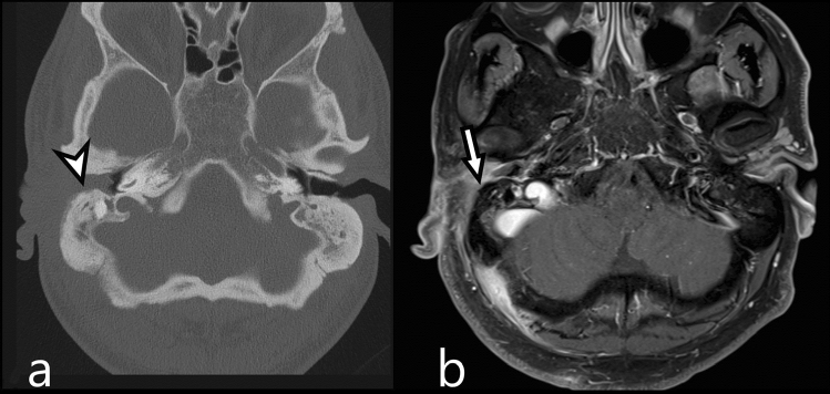
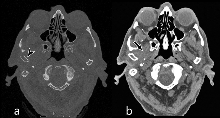
Necrotizing external otitis (NEO) is a serious complication of external otitis. NEO can be classified according to-anterior, medial, posterior, intracranial, and contralateral-extension patterns. Currently there is no consensus on the optimal imaging modality for the identification of disease extension. This study compares NEO extension patterns on MR and CT to evaluate diagnostic comparability.
Patients who received a CT and MR within a 3-month interval were retrospectively examined. Involvement of subsites and subsequent spreading patterns were assessed on both modalities by a radiologist in training and by a senior head and neck radiologist. The prevalence of extension patterns on CT and MR were calculated and compared.
All 21 included NEO cases showed an anterior extension pattern on CT and MR. Contrary to MR, medial extension was not recognized on CT in two out of six patients, and intracranial extension in five out of eight patients. The posterior extension pattern was not recognized on MR. Overall, single anterior extension pattern (62%) is more prevalent than multiple extension patterns (38%).
All anterior NEO extension pattern were identified on CT as well as MR. However, the medial and intracranial spreading patterns as seen on MR could only be identified on CT in a small number of patients. The posterior spreading pattern can be overlooked on MR. Thus, CT and MR are complimentary for the initial diagnosis and work-up of NEO as to correctly delineate disease extent through the skull base.
坏死性外耳道炎(NEO)是外耳道炎的严重并发症。根据前、中、后、颅内和对侧扩展模式,NEO 可进行分类。目前,对于识别疾病扩展的最佳成像方式尚未达成共识。本研究比较了 MR 和 CT 上 NEO 扩展模式,以评估诊断可比性。
回顾性检查了在 3 个月内接受 CT 和 MR 检查的患者。由一名受训放射科医生和一名资深头颈放射科医生在两种模式下评估亚部位受累情况和随后的扩散模式。计算并比较了 CT 和 MR 上的扩展模式的患病率。
所有 21 例纳入的 NEO 病例在 CT 和 MR 上均显示出前向扩展模式。与 MR 相反,6 例中有 2 例在 CT 上未识别出内侧扩展,8 例中有 5 例在 CT 上未识别出颅内扩展。MR 上未识别出后向扩展模式。总体而言,单一的前向扩展模式(62%)比多种扩展模式(38%)更为常见。
所有前向 NEO 扩展模式在 CT 和 MR 上均可识别。然而,MR 上所见的内侧和颅内扩展模式仅在少数患者的 CT 上可识别。MR 可能会忽略后向扩展模式。因此,CT 和 MR 可互补用于 NEO 的初始诊断和评估,以通过颅底正确描绘疾病范围。