van Kroonenburgh A M J L, van der Meer W L, Bothof R J P, van Tilburg M, van Tongeren J, Postma A A
1Department of Radiology, Maastricht University Medical Center, P. Debyelaan 25, 6229 HX Maastricht, The Netherlands.
2Department of Anesthesiology, Maastricht University Medical Center, P. Debyelaan 25, 6229 HX Maastricht, The Netherlands.
Curr Radiol Rep. 2018;6(1):3. doi: 10.1007/s40134-018-0263-y. Epub 2018 Jan 22.
To give an up-to-date overview of the strengths and weaknesses of current imaging modalities in diagnosis and follow-up of skull base osteomyelitis (SBO).
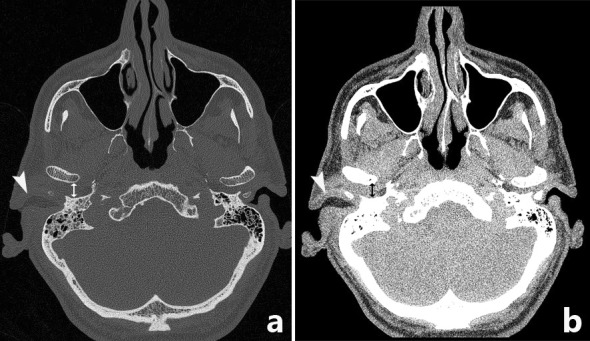
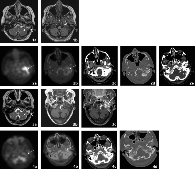
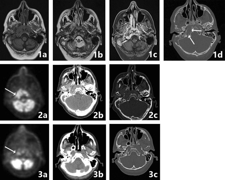
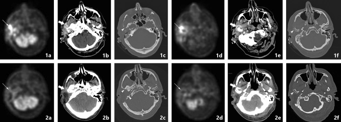
CT and MRI are both used for anatomical imaging, and nuclear techniques aid in functional process imaging. Hybrid techniques PET-CT and PET-MRI are the newest modalities which combine imaging strengths.
No single modality is able to address the scope of SBO. A combination of functional and anatomical imaging is needed, in the case of newly suspected SBO we suggest the use of PET-MRI (T1, T2, T1-FS-GADO, DWI) and separate HRCT for diagnosis and follow-up.
对当前成像方式在颅底骨髓炎(SBO)诊断及随访中的优缺点进行最新概述。
CT和MRI均用于解剖成像,核技术有助于功能过程成像。PET-CT和PET-MRI等混合技术是结合了成像优势的最新方式。
没有单一的成像方式能够全面解决SBO的问题。需要功能成像和解剖成像相结合,对于新怀疑的SBO病例,我们建议使用PET-MRI(T1、T2、T1-FS-GADO、DWI)和单独的高分辨率CT进行诊断和随访。