Department of Mechanical Engineering, Stanford University, Stanford, CA, USA.
Department of Electrical Engineering and Automation, Aalto University, Espoo, Finland.
Biomed Microdevices. 2021 Apr 26;23(2):27. doi: 10.1007/s10544-021-00547-2.
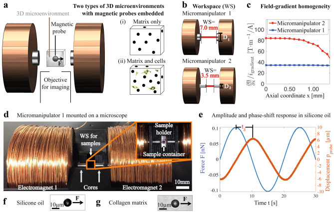
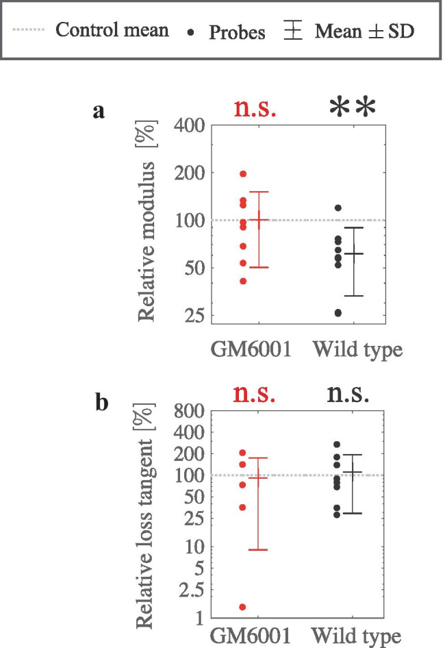
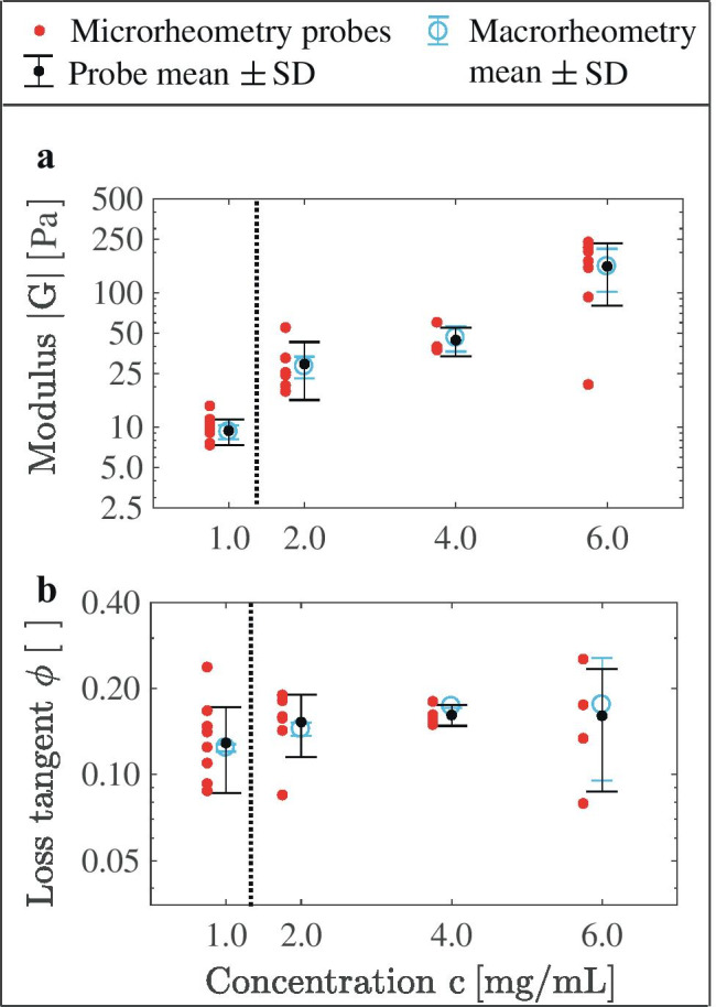
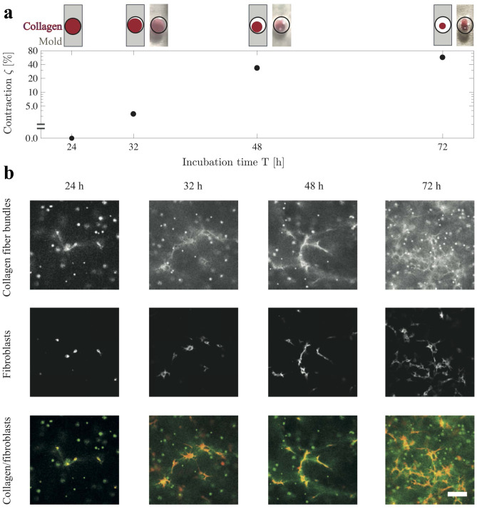
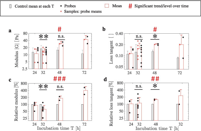
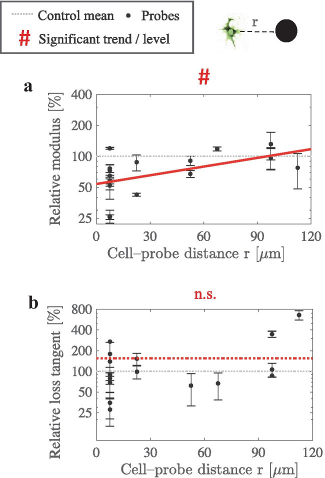
Changes in extracellular matrix stiffness impact a variety of biological processes including cancer progression. However, cells also actively remodel the matrices they interact with, dynamically altering the matrix mechanics they respond to. Further, cells not only react to matrix stiffness, but also have a distinct reaction to matrix viscoelasticity. The impact of cell-driven matrix remodeling on matrix stiffness and viscoelasticity at the microscale remains unclear, as existing methods to measure mechanics are largely at the bulk scale or probe only the surface of matrices, and focus on stiffness. Yet, establishing the impact of the matrix remodeling at the microscale is crucial to obtaining an understanding of mechanotransduction in biological matrices, and biological matrices are not just elastic, but are viscoelastic. Here, we advanced magnetic probe-based microrheology to overcome its previous limitations in measuring viscoelasticity at the cell-size-scale spatial resolution within 3D cell cultures that have tissue-relevant stiffness levels up to a Young's modulus of 0.5 kPa. Our magnetic microrheometers exert controlled magnetic forces on magnetic microprobes within reconstituted extracellular matrices and detect microprobe displacement responses to measure matrix viscoelasticity and determine the frequency-dependent shear modulus (stiffness), the loss tangent, and spatial heterogeneity. We applied these tools to investigate how microscale viscoelasticity of collagen matrices is altered by fibroblast cells as they contract collagen gels, a process studied extensively at the macroscale. Interestingly, we found that fibroblasts first soften the matrix locally over the first 32 hours of culture, and then progressively stiffen the matrix thereafter. Fibroblast activity also progressively increased the matrix loss tangent. We confirmed that the softening is caused by matrix-metalloproteinase-mediated collagen degradation, whereas stiffening is associated with local alignment and densification of collagen fibers around the fibroblasts. This work paves the way for the use of measurement systems that quantify microscale viscoelasticity within 3D cell cultures for studies of cell-matrix interactions in cancer progression and other areas.
细胞外基质硬度的变化会影响多种生物学过程,包括癌症进展。然而,细胞也会主动重塑它们相互作用的基质,动态改变它们所响应的基质力学。此外,细胞不仅对基质硬度有反应,而且对基质粘弹性也有明显的反应。细胞驱动的基质重塑对微尺度上基质硬度和粘弹性的影响尚不清楚,因为现有的力学测量方法在很大程度上是在体尺度上进行的,或者仅探测基质的表面,并且侧重于硬度。然而,确定微尺度上基质重塑的影响对于理解生物基质中的力学转导至关重要,而且生物基质不仅具有弹性,而且具有粘弹性。在这里,我们采用基于磁探针的微流变学方法来克服其在具有组织相关硬度水平(高达 0.5 kPa 的杨氏模量)的 3D 细胞培养中在细胞大小尺度空间分辨率下测量粘弹性的先前限制。我们的磁微流变仪在重构的细胞外基质内对磁性微探针施加受控的磁场力,并检测微探针的位移响应,以测量基质粘弹性并确定频率相关的剪切模量(硬度)、损耗角正切和空间异质性。我们应用这些工具来研究成纤维细胞收缩胶原凝胶时如何改变胶原基质的微尺度粘弹性,这一过程在宏观尺度上已经得到了广泛研究。有趣的是,我们发现成纤维细胞在培养的最初 32 小时内首先使基质局部变软,然后在此后逐渐使基质变硬。成纤维细胞的活性也逐渐增加了基质的损耗角正切。我们证实,软化是由基质金属蛋白酶介导的胶原降解引起的,而变硬与成纤维细胞周围胶原纤维的局部排列和致密化有关。这项工作为在癌症进展和其他领域的细胞-基质相互作用研究中使用量化 3D 细胞培养中微尺度粘弹性的测量系统铺平了道路。