Division of Radiology, Geneva University Hospitals, Chemin du Pont-Bochet 3, 1226, Thonex, Switzerland.
Image Guided Interventions Laboratory, University of Geneva, Rue Gabrielle-Perret-Gentil 4, 1205, Geneva, Switzerland.
Neuroradiology. 2021 Sep;63(9):1569-1573. doi: 10.1007/s00234-021-02717-8. Epub 2021 Apr 28.
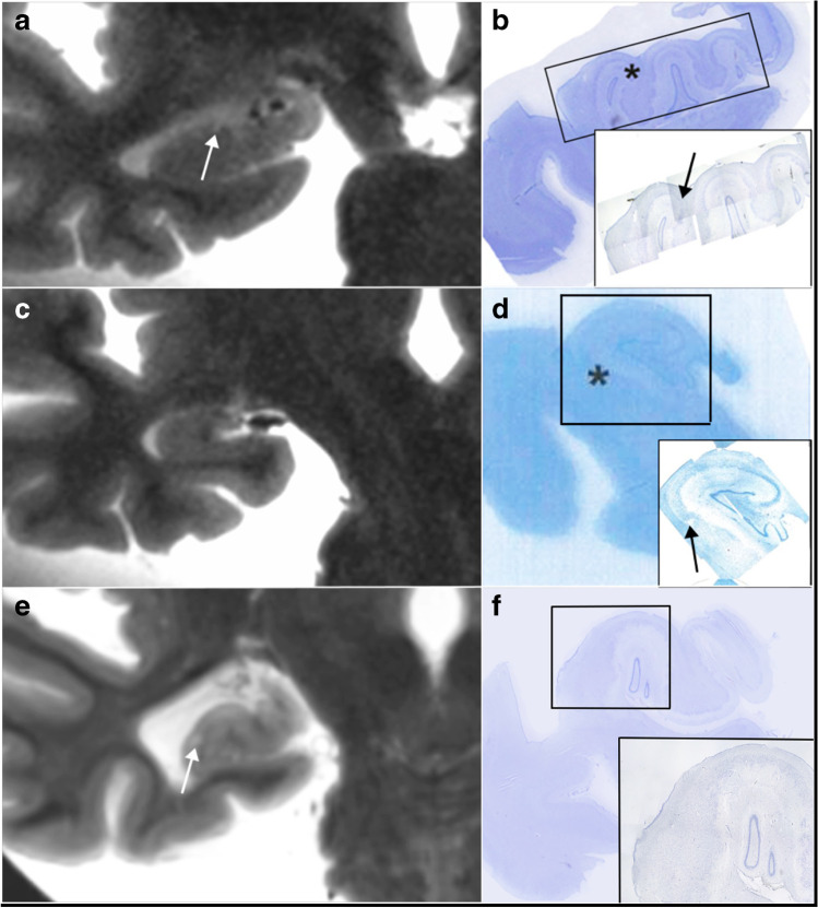
Cortical microinfarcts (CMI) are increasingly recognized in the neurological community as a biomarker related to cognitive impairment and dementia. If their radiological depiction has been largely described in experimental settings using ultra-high-field magnetic resonance imaging (MRI), less is known about their visibility on routinely used 3-T MRI. In this radiologic-pathologic correlation study, using 3-T post-mortem MRI, we searched for hippocampal CMI, in a double-blinded fashion, and found that only 4/36, or 11%, were clearly demonstrated on both radiological and histopathological exams.
皮质微梗死(CMI)在神经科学界越来越被认为是与认知障碍和痴呆相关的生物标志物。虽然它们的影像学表现已经在实验环境中使用超高场磁共振成像(MRI)得到了广泛描述,但在常规使用的 3-T MRI 上的可见性知之甚少。在这项放射病理相关性研究中,我们使用 3-T 死后 MRI 进行双盲搜索,发现只有 4/36,即 11%,在影像学和组织病理学检查中都能明确显示。