Mukhtar Areej Hussein, Alqutub Montaser N
Department of Periodontics and Community Dentistry, College of Dentistry, King Saud University, Riyadh, Saudi Arabia.
Int J Appl Basic Med Res. 2021 Apr-Jun;11(2):75-79. doi: 10.4103/ijabmr.IJABMR_363_20. Epub 2021 Apr 8.
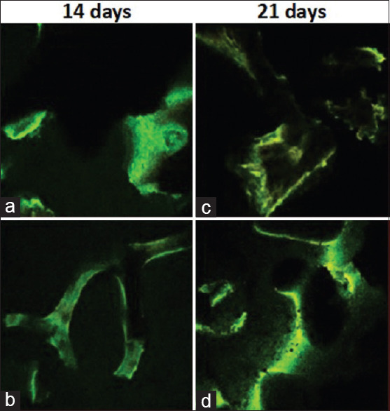
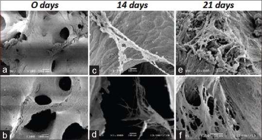
The aim of this study was to compare between equine and human bone blocks in the osteogenic differentiation of cultured human periodontal ligament stem cells (hPDLSCs) at 14 and 21 days of culture, using confocal laser microscopy and scanning electron microscopy.
cultures of commercially obtained hPDLSCs were seeded onto equine and human bone blocks. At 14 days and 21 days of culture, confocal laser microscope images were obtained to assess cellular differentiation and adhesion, and scanning electron microscope images were obtained to validate the osteogenic differentiation by showing the morphological characteristics of the new bone cells.
Both equine and human bone blocks showed positive staining for newly formed bone cells through the confocal laser microscope analysis, however, a higher signal intensity was expressed at 21 days of culture. These findings indicate the biocompatibility of hPDLSC with both types of bone blocks, cellular differentiation, and adhesion. Scanning electron microscopy images validated the osteogenic differentiation by showing the common characteristics of bone cells as flattened, polygonal morphology with multiple extending cytoplasmic processes.
Both equine and human bone blocks were able to confirm the osteogenic capability of seeded human PDLSC. There was no significant difference between equine and human bone blocks on the human PDLSC differentiation. Superior osteogenic differentiation of cultured hPDLSCs was evident at 21 days in comparison to 14 days.
本研究旨在利用共聚焦激光显微镜和扫描电子显微镜,比较马骨块和人骨块在培养14天和21天时对培养的人牙周膜干细胞(hPDLSCs)成骨分化的影响。
将商业获取的hPDLSCs培养物接种到马骨块和人骨块上。在培养14天和21天时,获取共聚焦激光显微镜图像以评估细胞分化和黏附情况,并获取扫描电子显微镜图像,通过显示新骨细胞的形态特征来验证成骨分化。
通过共聚焦激光显微镜分析,马骨块和人骨块对新形成的骨细胞均显示阳性染色,然而,在培养21天时表达的信号强度更高。这些发现表明hPDLSC与两种类型骨块具有生物相容性、细胞分化和黏附能力。扫描电子显微镜图像通过显示骨细胞扁平、多边形形态及多个延伸的细胞质突起的共同特征,验证了成骨分化。
马骨块和人骨块均能证实接种的人PDLSC的成骨能力。马骨块和人骨块对人PDLSC分化的影响无显著差异。与14天相比,培养的hPDLSCs在21天时成骨分化更明显。