• 文献检索
  • 文档翻译
  • 深度研究
  • 学术资讯
  • Suppr Zotero 插件Zotero 插件
  • 邀请有礼
  • 套餐&价格
  • 历史记录
应用&插件
Suppr Zotero 插件Zotero 插件浏览器插件Mac 客户端Windows 客户端微信小程序
定价
高级版会员购买积分包购买API积分包
服务
文献检索文档翻译深度研究API 文档MCP 服务
关于我们
关于 Suppr公司介绍联系我们用户协议隐私条款
关注我们

Suppr 超能文献

核心技术专利:CN118964589B侵权必究
粤ICP备2023148730 号-1Suppr @ 2026

文献检索

告别复杂PubMed语法,用中文像聊天一样搜索,搜遍4000万医学文献。AI智能推荐,让科研检索更轻松。

立即免费搜索

文件翻译

保留排版,准确专业,支持PDF/Word/PPT等文件格式,支持 12+语言互译。

免费翻译文档

深度研究

AI帮你快速写综述,25分钟生成高质量综述,智能提取关键信息,辅助科研写作。

立即免费体验

彩色双功能超声及计算机断层纳米造影剂分析巨细胞动脉炎裸鼠模型的发病机制

Analysis of Development Mechanism of Giant Cell Arteritis in Nude Mouse Model through Color Duplex Sonography and Computerized Tomography Nanocontrast Agent.

机构信息

Department of Radiology, The Second Affiliated Hospital of Dalian Medical University, Dalian 116031, China.

Department of Ultrasound, The Second Affiliated Hospital of Dalian Medical University, Dalian 116031, China.

出版信息

Biomed Res Int. 2021 Apr 9;2021:6627925. doi: 10.1155/2021/6627925. eCollection 2021.

DOI:10.1155/2021/6627925
PMID:33928153
原文链接:https://pmc.ncbi.nlm.nih.gov/articles/PMC8053047/
Abstract

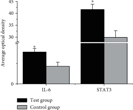
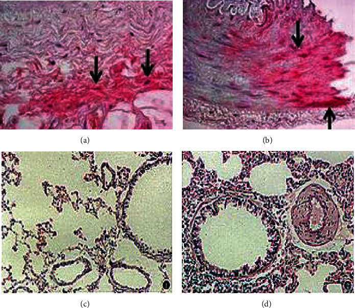
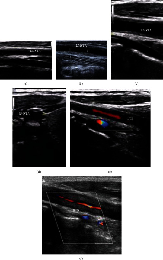
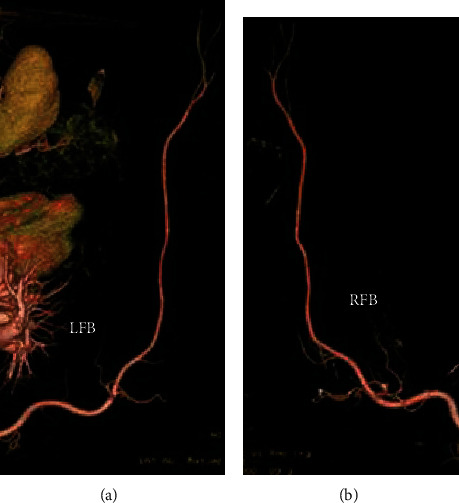
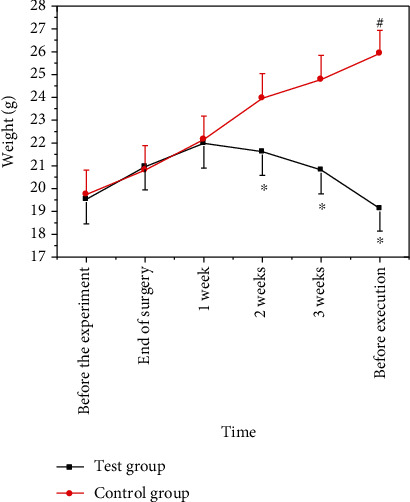
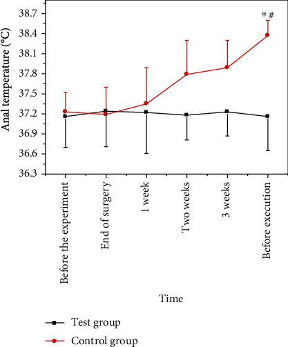
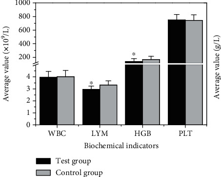
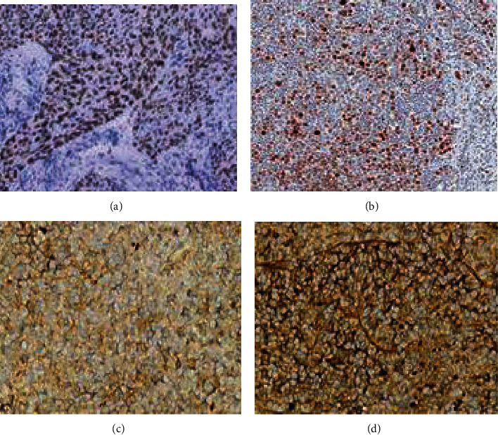
To explore the application value of color duplex sonography and enhanced computerized tomography (CT) inspection based on a nanocontrast agent in diagnosis and pathogenesis in giant cell arteritis (GCA), the GCA nude mouse model was constructed. In this study, 40 healthy male BalB/c nude mice aged 6-8 weeks were randomly divided into a control group (no model) and an experimental group (model), with 20 mice in each group, and the temporal artery tissue of GCA patients diagnosed as positive by temporal artery biopsy was implanted into nude mice to construct a GCA nude mouse model. Abdominal aortic biopsy and immunohistochemistry were used to verify the success of the GCA nude mouse model. All nude mice were subjected to color duplex sonography and enhanced CT examination based on a nanocontrast agent. At the same time, the basic indicators such as body weight, temperature, white blood cell (WBC), lymphocytes (LYM), hemoglobin (HGB), and platelet (PLT) were measured, and the protein expression levels of interleukin-6 (IL-6) and signal transducer and activator of transcription 3 (STAT3) were detected by immunohistochemistry. The results showed that the temporal artery wall of the nude mice in the experimental group thickened and the lumen was significantly narrowed, indicating that the cell arteritis model of nude mice was successfully constructed; ultrasound examination showed that the right superficial temporal artery vascular cavity narrowed, the blood flow signal changed like a filling defect around the periphery, and there was a low echo halo. CT examination showed that the left superficial temporal artery narrowed, and the inner diameter of the narrow segment of blood vessels changed like a bead. The body weight of nude mice in the experimental group decreased significantly after the modeling was completed ( < 0.05); after modeling, the body temperature of the nude mice in the experimental group increased significantly ( < 0.05); LYM and HGB values of nude mice in the experimental group were significantly lower than those in the control group ( < 0.05); the content of IL-6, STAT3, IL-6, and STAT3 proteins in the arterial tissue of nude mice in the experimental group was lower than that of the control group ( < 0.05), indicating that color duplex sonography and CT contrast agent technology can be used in the diagnosis and development mechanism research of GC.

摘要

为了探讨基于纳米造影剂的彩色双功能超声与增强计算机断层扫描(CT)检查在巨细胞动脉炎(GCA)诊断和发病机制中的应用价值,构建了 GCA 裸鼠模型。本研究中,将 40 只 6-8 周龄的健康雄性 BalB/c 裸鼠随机分为对照组(无模型)和实验组(模型),每组 20 只,将经颞动脉活检诊断为阳性的 GCA 患者的颞动脉组织植入裸鼠体内构建 GCA 裸鼠模型。腹主动脉活检和免疫组织化学用于验证 GCA 裸鼠模型的成功构建。所有裸鼠均接受基于纳米造影剂的彩色双功能超声和增强 CT 检查。同时,测量裸鼠的体重、体温、白细胞(WBC)、淋巴细胞(LYM)、血红蛋白(HGB)和血小板(PLT)等基本指标,并通过免疫组织化学检测白细胞介素-6(IL-6)和信号转导与转录激活因子 3(STAT3)的蛋白表达水平。结果显示,实验组裸鼠的颞动脉壁增厚,管腔明显变窄,表明裸鼠细胞动脉炎模型构建成功;超声检查显示右侧颞浅动脉血管腔变窄,血流信号周边呈充盈缺损样改变,并有低回声晕;CT 检查显示左侧颞浅动脉变窄,血管狭窄段内径呈串珠样改变。建模完成后,实验组裸鼠体重明显下降(<0.05);建模后,实验组裸鼠体温明显升高(<0.05);实验组裸鼠 LYM 和 HGB 值明显低于对照组(<0.05);实验组裸鼠动脉组织中 IL-6、STAT3、IL-6 和 STAT3 蛋白含量均低于对照组(<0.05),表明彩色双功能超声与 CT 造影剂技术可用于 GCA 的诊断和发病机制研究。

https://cdn.ncbi.nlm.nih.gov/pmc/blobs/6b43/8053047/162bf4eed882/BMRI2021-6627925.009.jpg
https://cdn.ncbi.nlm.nih.gov/pmc/blobs/6b43/8053047/d86a33575a71/BMRI2021-6627925.001.jpg
https://cdn.ncbi.nlm.nih.gov/pmc/blobs/6b43/8053047/c9338892e57a/BMRI2021-6627925.002.jpg
https://cdn.ncbi.nlm.nih.gov/pmc/blobs/6b43/8053047/2ecd6ce66be9/BMRI2021-6627925.003.jpg
https://cdn.ncbi.nlm.nih.gov/pmc/blobs/6b43/8053047/d920b0bddc12/BMRI2021-6627925.004.jpg
https://cdn.ncbi.nlm.nih.gov/pmc/blobs/6b43/8053047/ed6c0b4f9be3/BMRI2021-6627925.005.jpg
https://cdn.ncbi.nlm.nih.gov/pmc/blobs/6b43/8053047/b47f7c22786a/BMRI2021-6627925.006.jpg
https://cdn.ncbi.nlm.nih.gov/pmc/blobs/6b43/8053047/370424654ca1/BMRI2021-6627925.007.jpg
https://cdn.ncbi.nlm.nih.gov/pmc/blobs/6b43/8053047/716ddd5199ee/BMRI2021-6627925.008.jpg
https://cdn.ncbi.nlm.nih.gov/pmc/blobs/6b43/8053047/162bf4eed882/BMRI2021-6627925.009.jpg

相似文献

1
Analysis of Development Mechanism of Giant Cell Arteritis in Nude Mouse Model through Color Duplex Sonography and Computerized Tomography Nanocontrast Agent.彩色双功能超声及计算机断层纳米造影剂分析巨细胞动脉炎裸鼠模型的发病机制
Biomed Res Int. 2021 Apr 9;2021:6627925. doi: 10.1155/2021/6627925. eCollection 2021.
2
Colour duplex sonography of temporal arteries before decision for biopsy: a prospective study in 55 patients with suspected giant cell arteritis.活检决策前颞动脉的彩色双功超声检查:55例疑似巨细胞动脉炎患者的前瞻性研究
Arthritis Res Ther. 2006;8(4):R116. doi: 10.1186/ar2003.
3
[Value of color-coded duplex ultrasound in patients with polymyalgia rheumatica without signs of temporal arteritis].[彩色编码双功超声在无颞动脉炎体征的风湿性多肌痛患者中的价值]
Dtsch Med Wochenschr. 2000 Oct 20;125(42):1250-6. doi: 10.1055/s-2000-7853.
4
The role of color duplex sonography in the diagnosis of giant cell arteritis.彩色双功超声在巨细胞动脉炎诊断中的作用。
J Ultrasound Med. 2004 Nov;23(11):1493-8. doi: 10.7863/jum.2004.23.11.1493.
5
Comparison between color duplex ultrasonography and histology of the temporal artery in cranial arteritis (giant cell arteritis).颅动脉炎(巨细胞动脉炎)颞动脉的彩色双功能超声检查与组织学比较
Eur J Med Res. 2003 Jan 28;8(1):1-7.
6
Clinical and color Doppler imaging features of one patient with occult giant cell arteritis presenting arteritic anterior ischemic optic neuropathy.一名隐匿性巨细胞动脉炎患者出现动脉炎性前部缺血性视神经病变的临床及彩色多普勒成像特征。
Rom J Morphol Embryol. 2016;57(2):579-83.
7
The predictive value of the halo sign in color Doppler ultrasonography of the temporal arteries for diagnosing giant cell arteritis.颞动脉彩色多普勒超声检查中晕征对诊断巨细胞动脉炎的预测价值。
J Rheumatol. 2002 Jun;29(6):1224-6.
8
Clinical, Laboratory and Ultrasonographic Interrelations in Giant Cell Arteritis.巨细胞动脉炎的临床、实验室和超声表现的相关性。
J Stroke Cerebrovasc Dis. 2021 Apr;30(4):105601. doi: 10.1016/j.jstrokecerebrovasdis.2021.105601. Epub 2021 Jan 23.
9
Bilateral axillobrachial and external carotid artery manifestation of giant cell arteritis: important role of color duplex ultrasonography in the diagnosis.
Int Angiol. 2005 Jun;24(2):202-5.
10
[Use of high resolution color Doppler sonography in diagnosis of temporal arteritis].高分辨率彩色多普勒超声在颞动脉炎诊断中的应用
Rofo. 1998 Dec;169(6):605-8. doi: 10.1055/s-2007-1015349.

引用本文的文献

1
Retracted: Analysis of Development Mechanism of Giant Cell Arteritis in Nude Mouse Model through Color Duplex Sonography and Computerized Tomography Nanocontrast Agent.
Biomed Res Int. 2024 Jan 9;2024:9762671. doi: 10.1155/2024/9762671. eCollection 2024.
2
Animal models for large vessel vasculitis - The unmet need.大血管血管炎的动物模型——未满足的需求。
Rheumatol Immunol Res. 2023 Apr 18;4(1):4-10. doi: 10.2478/rir-2023-0002. eCollection 2023 Mar.

本文引用的文献

1
Temporal artery biopsy versus imaging in patients with cranial giant cell arteritis.颞动脉活检与影像学检查在颅巨人细胞动脉炎患者中的应用比较。
Radiol Med. 2020 Sep;125(9):902-903. doi: 10.1007/s11547-020-01166-2. Epub 2020 Mar 19.
2
Lessons of the month 4: Giant cell arteritis with normal inflammatory markers and isolated oculomotor nerve palsy.本月病例 4:炎症指标正常的巨细胞动脉炎伴孤立性动眼神经麻痹。
Clin Med (Lond). 2020 Mar;20(2):224-226. doi: 10.7861/clinmed.2019-0504.
3
Novel ultrasonographic Halo Score for giant cell arteritis: assessment of diagnostic accuracy and association with ocular ischaemia.
新型巨细胞动脉炎超声 Halo 评分:诊断准确性评估及其与眼部缺血的关系。
Ann Rheum Dis. 2020 Mar;79(3):393-399. doi: 10.1136/annrheumdis-2019-216343. Epub 2020 Jan 3.
4
Clinical symptoms and associated vascular imaging findings in Takayasu's arteritis compared to giant cell arteritis.与巨细胞动脉炎相比,Takayasu 动脉炎的临床症状及相关血管影像学表现。
Ann Rheum Dis. 2020 Feb;79(2):262-267. doi: 10.1136/annrheumdis-2019-216145. Epub 2019 Oct 24.
5
Radiation Exposure During Transarterial Chemoembolization: Angio-CT Versus Cone-Beam CT.经动脉化疗栓塞术中的辐射暴露:血管造影 CT 与锥形束 CT。
Cardiovasc Intervent Radiol. 2019 Nov;42(11):1609-1618. doi: 10.1007/s00270-019-02269-8. Epub 2019 Jun 20.
6
[Current Recommendations for Diagnostics in Giant Cell Arteritis].[巨细胞动脉炎诊断的当前建议]
Dtsch Med Wochenschr. 2019 May;144(9):587-594. doi: 10.1055/a-0831-0812. Epub 2019 Apr 26.
7
Factors Associated with Relapse and Dependence on Glucocorticoids in Giant Cell Arteritis.巨细胞动脉炎复发和对糖皮质激素依赖的相关因素。
J Rheumatol. 2020 Jan;47(1):108-116. doi: 10.3899/jrheum.181127. Epub 2019 Mar 15.
8
Nanoliposomes Co-Encapsulating CT Imaging Contrast Agent and Photosensitizer for Enhanced, Imaging Guided Photodynamic Therapy of Cancer.载 CT 成像造影剂和光敏剂的纳米脂质体用于增强癌症的成像引导光动力治疗。
Theranostics. 2019 Feb 12;9(5):1323-1335. doi: 10.7150/thno.31079. eCollection 2019.
9
Cytokines, growth factors and proteases in medium and large vessel vasculitis.中大型血管炎中的细胞因子、生长因子和蛋白酶。
Clin Immunol. 2019 Sep;206:33-41. doi: 10.1016/j.clim.2019.02.007. Epub 2019 Feb 14.
10
Diagnostic performance of temporal artery ultrasound for the diagnosis of giant cell arteritis: a systematic review and meta-analysis of the literature.颞动脉超声对巨细胞动脉炎诊断的诊断性能:文献的系统评价和荟萃分析。
Autoimmun Rev. 2019 Jan;18(1):56-61. doi: 10.1016/j.autrev.2018.07.012. Epub 2018 Nov 5.