Department of Radiology, Leiden University Medical Center (LUMC), Albinusdreef 2, 2333, ZA, Leiden, The Netherlands.
Department of Radiology, University Medical Center Utrecht, Heidelberglaan 100, 3584, CX, Utrecht, The Netherlands.
Eur Radiol. 2021 Nov;31(11):8208-8217. doi: 10.1007/s00330-021-07970-2. Epub 2021 Apr 30.
The underlying structural brain correlates of neuropsychiatric involvement in systemic lupus erythematosus (NPSLE) remain unclear, thus hindering correct diagnosis. We compared brain tissue volumes between a clinically well-defined cohort of patients with NPSLE and SLE patients with neuropsychiatric syndromes not attributed to SLE (non-NPSLE). Within the NPSLE patients, we also examined differences between patients with two distinct disease phenotypes: ischemic and inflammatory.
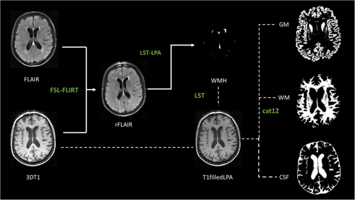
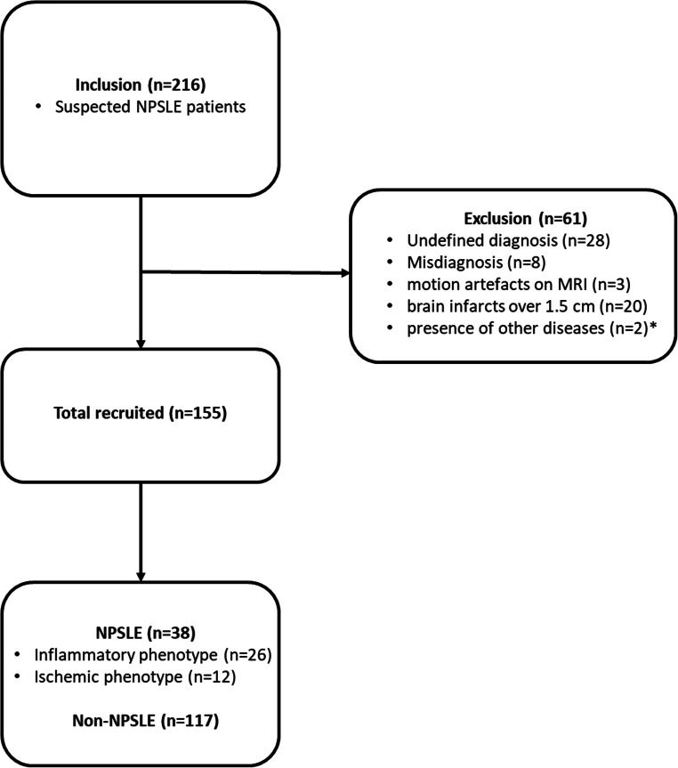
In this prospective (May 2007 to April 2015) cohort study, we included 38 NPSLE patients (26 inflammatory and 12 ischemic) and 117 non-NPSLE patients. All patients underwent a 3-T brain MRI scan that was used to automatically determine white matter, grey matter, white matter hyperintensities (WMH) and total brain volumes. Group differences in brain tissue volumes were studied with linear regression analyses corrected for age, gender, and total intracranial volume and expressed as B values and 95% confidence intervals.
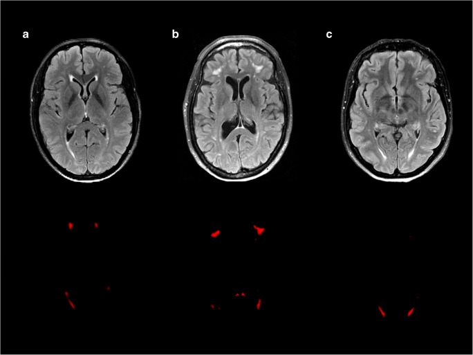
NPSLE patients showed higher WMH volume compared to non-NPSLE patients (p = 0.004). NPSLE inflammatory patients showed lower total brain (p = 0.014) and white matter volumes (p = 0.020), and higher WMH volume (p = 0.002) compared to non-NPSLE patients. Additionally, NPSLE inflammatory patients showed lower white matter (p = 0.020) and total brain volumes (p = 0.038) compared to NPSLE ischemic patients.
We showed that different phenotypes of NPSLE were related to distinct patterns of underlying structural brain MRI changes. Especially the inflammatory phenotype of NPSLE was associated with the most pronounced brain volume changes, which might facilitate the diagnostic process in SLE patients with neuropsychiatric symptoms.
• Neuropsychiatric systemic lupus erythematosus (NPSLE) patients showed a higher WMH volume compared to SLE patients with neuropsychiatric syndromes not attributed to SLE (non-NPSLE). • NPSLE patients with inflammatory phenotype showed a lower total brain and white matter volume, and a higher volume of white matter hyperintensities, compared to non-NPSLE patients. • NPSLE patients with inflammatory phenotype showed lower white matter and total brain volumes compared to NPSLE patients with ischemic phenotype.
系统性红斑狼疮(SLE)伴神经精神性狼疮(NPSLE)患者的潜在结构性脑相关因素仍不清楚,这阻碍了正确的诊断。我们比较了一组临床明确的 NPSLE 患者和 SLE 伴非 SLE 神经精神综合征(non-NPSLE)患者的脑组织体积。在 NPSLE 患者中,我们还检查了两种不同疾病表型(缺血性和炎症性)之间的差异。
本前瞻性队列研究(2007 年 5 月至 2015 年 4 月)纳入 38 例 NPSLE 患者(26 例炎症性和 12 例缺血性)和 117 例 non-NPSLE 患者。所有患者均行 3T 脑 MRI 扫描,用于自动确定脑白质、灰质、脑白质高信号(WMH)和总脑容量。通过线性回归分析,对年龄、性别和总颅内体积进行校正,并以 B 值和 95%置信区间表示,比较脑组织结构体积的组间差异。
与 non-NPSLE 患者相比,NPSLE 患者的 WMH 体积更高(p=0.004)。与 non-NPSLE 患者相比,NPSLE 炎症性患者的总脑(p=0.014)和白质体积(p=0.020)更低,WMH 体积更高(p=0.002)。此外,与 NPSLE 缺血性患者相比,NPSLE 炎症性患者的白质(p=0.020)和总脑体积(p=0.038)更低。
我们发现,NPSLE 的不同表型与潜在的结构性脑 MRI 变化模式有关。特别是 NPSLE 的炎症表型与最明显的脑体积变化有关,这可能有助于 SLE 伴神经精神症状患者的诊断过程。
NPSLE 患者的 WMH 体积高于 non-NPSLE 患者。
与 non-NPSLE 患者相比,NPSLE 炎症性患者的总脑和白质体积更低,WMH 体积更高。
与 NPSLE 缺血性患者相比,NPSLE 炎症性患者的白质和总脑体积更低。