Alaoui Ahmed Amine, Oumedjbeur Kussil, Djinbachian Roupen, Marchand Étienne, Marques Paola N, Bouin Mickael, Bouchard Simon, von Renteln Daniel
University of Montreal, Faculty of Medicine, Montreal, QC, Canada.
University of Montreal Hospital Centre Research Center, Gastroenterology, Montreal, QC, Canada.
Endosc Int Open. 2021 May;9(5):E684-E692. doi: 10.1055/a-1388-6694. Epub 2021 Apr 22.
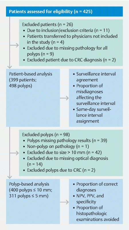
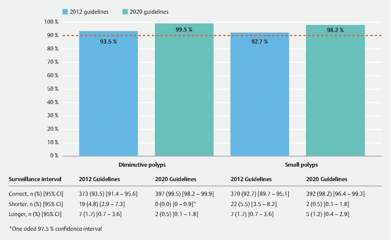
A novel endoscopic optical diagnosis classification system (SIMPLE) has recently been developed. This study aimed to evaluate the SIMPLE classification in a clinical cohort. All diminutive and small colorectal polyps found in a cohort of individuals undergoing screening, diagnostic, or surveillance colonoscopies underwent optical diagnosis using image-enhanced endoscopy (IEE) and the SIMPLE classification. The primary outcome was the agreement of surveillance intervals determined by optical diagnosis compared with pathology-based results for diminutive polyps. Secondary outcomes included the negative predictive value (NPV) for rectosigmoid adenomas, the percentage of pathology exams avoided, and the percentage of immediate surveillance interval recommendations. Analysis of optical diagnosis for polyps ≤ 10 mm was also performed. 399 patients (median age 62.6 years; 55.6 % female) were enrolled. For patients with at least one polyp ≤ 5 mm undergoing optical diagnosis, agreement with pathology-based surveillance intervals was 93.5 % (95 % confidence interval [CI] 91.4-95.6). The NPV for rectosigmoid adenomas was 86.7 % (95 %CI 77.5-93.2). When using optical diagnosis, pathology analysis could be avoided in 61.5 % (95 %CI 56.9-66.2) of diminutive polyps, and post-colonoscopy surveillance intervals could be given immediately to 70.9 % (95 %CI 66.5-75.4) of patients. For patients with at least one ≤ 10 mm polyp, agreement with pathology-based surveillance intervals was 92.7 % (95 %CI 89.7-95.1). NPV for rectosigmoid adenomas ≤ 10 mm was 85.1 % (95 %CI CI 76.3-91.6). IEE with the SIMPLE classification achieved the quality benchmark for the resect and discard strategy; however, the NPV for rectosigmoid polyps requires improvement.
一种新型的内镜光学诊断分类系统(SIMPLE)最近已被开发出来。本研究旨在评估临床队列中的SIMPLE分类。在接受筛查、诊断或监测结肠镜检查的个体队列中发现的所有微小和小的结直肠息肉,均采用图像增强内镜检查(IEE)和SIMPLE分类进行光学诊断。主要结局是光学诊断确定的监测间隔与微小息肉基于病理结果的一致性。次要结局包括直肠乙状结肠腺瘤的阴性预测值(NPV)、避免的病理检查百分比以及立即监测间隔建议的百分比。还对≤10mm息肉的光学诊断进行了分析。共纳入399例患者(中位年龄62.6岁;55.6%为女性)。对于至少有一个≤5mm息肉接受光学诊断的患者,与基于病理的监测间隔的一致性为93.5%(95%置信区间[CI]91.4 - 95.6)。直肠乙状结肠腺瘤的NPV为86.7%(95%CI 77.5 - 93.2)。使用光学诊断时,61.5%(95%CI 56.9 - 66.2)的微小息肉可避免病理分析,70.9%(95%CI 66.5 - 75.4)的患者在结肠镜检查后可立即获得监测间隔。对于至少有一个≤10mm息肉的患者,与基于病理的监测间隔的一致性为92.7%(95%CI 89.7 - 95.1)。≤10mm直肠乙状结肠腺瘤的NPV为85.1%(95%CI 76.3 - 91.6)。采用SIMPLE分类的IEE达到了切除并丢弃策略的质量基准;然而,直肠乙状结肠息肉的NPV需要改进。