Third Hospital Orthopaedics Department, Beijing Key Laboratory of Spinal Disease Research, Peking University, Beijing, China.
Peking University Health Science Center, Beijing, China.
Orthop Surg. 2021 Jun;13(4):1141-1148. doi: 10.1111/os.12962. Epub 2021 May 4.
Numerous studies have applied a variety of methods to assess paraspinal muscle degeneration. However, the methodological differences in imaging evaluation may lead to imprecise or inconsistent results. This article aimed to provide a pragmatic summary review of the current imaging modalities, measurement protocols, and imaging parameters in the evaluation of paraspinal muscle fat infiltration (FI) in MRI studies.
Web of Science, EMBASE, and PubMed were searched from January 2005 to March 2020 to identify studies that examined the FI of paraspinal muscles on MRI among patients with lumbar degenerative diseases.
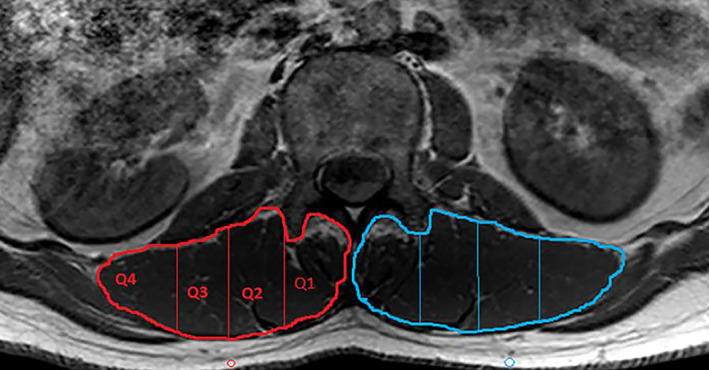
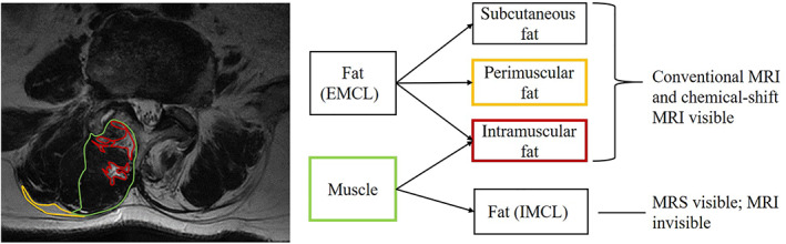
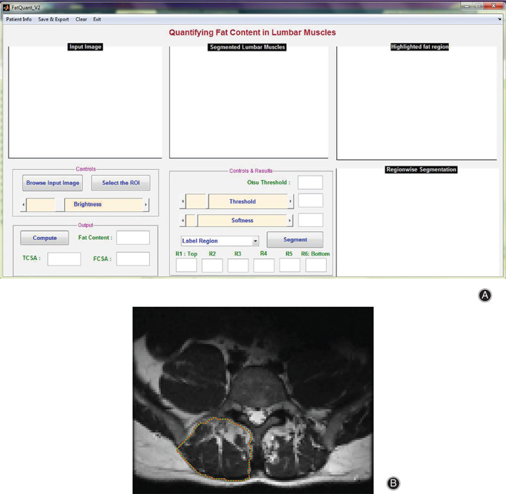
Intramyocellular lipids measured by magnetic resonance spectroscopy and FI measured by chemical-shift MRI were both correlated to low back pain and several degenerative lumbar diseases, whereas results on the relationship between FI and degenerative lumbar pathologies using conventional MRI were conflicting. Multi-segment measurement of FI at the lesion segment and adjacent segments could be a prognostic indicator for lumbar surgery. Most studies adopted the center of the intervertebral disc or endplate as the level of slice to evaluate the FI. Compared with visual semiquantitative assessment, quantitative parameters appeared to be precise for eliminating individual or modality differences. It has been demonstrated that fat CSA/total CSA (based on area) and muscle-fat index (based on signal intensity) as quantitative FI parameters are associated with multiple lumbar diseases and clinical outcomes after surgery.
Having a good command of the methodology of paraspinal muscle FI on MRI was effective for diagnosis and prognosis in clinical practice.
许多研究采用了多种方法来评估脊柱旁肌肉退变。然而,影像学评估方法的差异可能导致结果不精确或不一致。本文旨在提供一个实用的综述,总结目前 MRI 研究中评估脊柱旁肌肉脂肪浸润(FI)的各种成像方式、测量方案和成像参数。
检索 2005 年 1 月至 2020 年 3 月的 Web of Science、EMBASE 和 PubMed 数据库,以确定评估腰椎退行性疾病患者脊柱旁肌肉 FI 的 MRI 研究。
磁共振波谱测量的肌细胞内脂质和化学位移 MRI 测量的 FI 均与腰痛和几种退行性腰椎疾病相关,而常规 MRI 上 FI 与退行性腰椎病变之间的关系结果存在争议。病变节段和相邻节段的 FI 多节段测量可能是腰椎手术的预后指标。大多数研究采用椎间盘中心或终板作为切片评估 FI 的水平。与视觉半定量评估相比,定量参数似乎更能精确消除个体或模态差异。已经证明,基于面积的脂肪 CSA/总 CSA(基于面积)和基于信号强度的肌肉脂肪指数作为定量 FI 参数与多种腰椎疾病和术后临床结果相关。
熟练掌握 MRI 上脊柱旁肌肉 FI 的方法学对临床实践中的诊断和预后具有重要意义。