Department of Clinical Sciences, Polytechnic University of Marche, 60131 Ancona, Italy.
Nutrition and Food Science Group, Department of Analytical and Food Chemistry, CITACA, CACTI, University of Vigo, 36310 Vigo, Spain.
Molecules. 2021 Apr 29;26(9):2615. doi: 10.3390/molecules26092615.
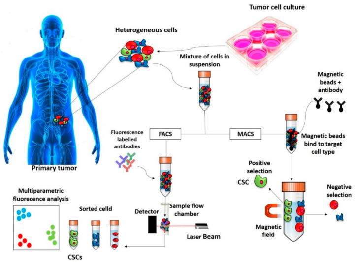
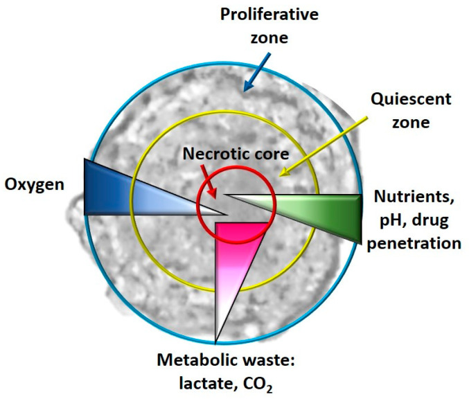
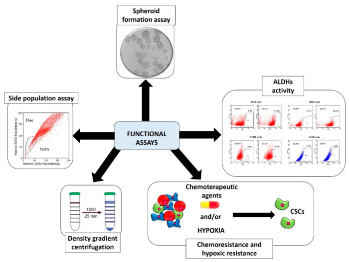
Cancer stem cells (CSCs) are a rare tumor subpopulation with high differentiation, proliferative and tumorigenic potential compared to the remaining tumor population. CSCs were first discovered by Bonnet and Dick in 1997 in acute myeloid leukemia. The identification and isolation of these cells in this pioneering study were carried out through the flow cytometry, exploiting the presence of specific cell surface molecular markers (CD34/CD38). In the following years, different strategies and projects have been developed for the study of CSCs, which are basically divided into surface markers assays and functional assays; some of these techniques also allow working with a cellular model that better mimics the tumor architecture. The purpose of this mini review is to summarize and briefly describe all the current methods used for the identification, isolation and enrichment of CSCs, describing, where possible, the molecular basis, the advantages and disadvantages of each technique with a particular focus on those that offer a three-dimensional culture.
癌症干细胞(CSC)是一种罕见的肿瘤亚群,与剩余的肿瘤群体相比,其具有高分化、增殖和致瘤潜能。Bonnet 和 Dick 于 1997 年在急性髓系白血病中首次发现了 CSC。在这项开创性的研究中,通过流式细胞术对这些细胞进行了鉴定和分离,利用了特定的细胞表面分子标记(CD34/CD38)的存在。在随后的几年中,为了研究 CSC,已经开发出了不同的策略和项目,这些策略基本上分为表面标记物分析和功能分析;其中一些技术还允许使用更能模拟肿瘤结构的细胞模型。本综述的目的是总结和简要描述目前用于鉴定、分离和富集 CSC 的所有方法,尽可能描述每种技术的分子基础、优缺点,特别关注那些提供三维培养的技术。