Schneider Felix, Duong Thuy-An, Rust Marco B
Molecular Neurobiology Group, Institute of Physiological Chemistry, University of Marburg, Marburg 35032, Germany.
Center for Mind, Brain and Behavior (CMBB), University of Marburg and Justus-Liebig-University Giessen, Marburg 35032, Germany.
eNeuro. 2021 May 20;8(3). doi: 10.1523/ENEURO.0536-20.2021. Print 2021 May-Jun.
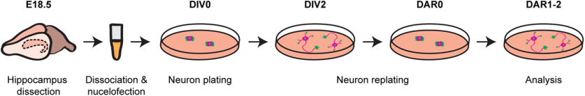
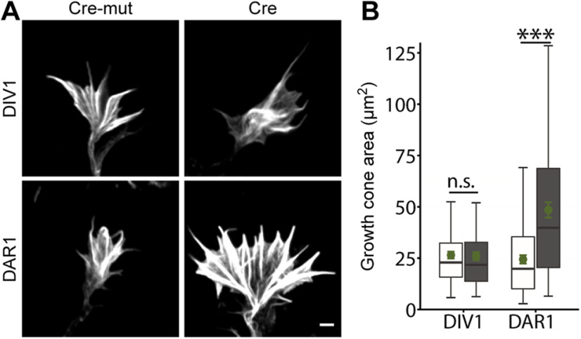
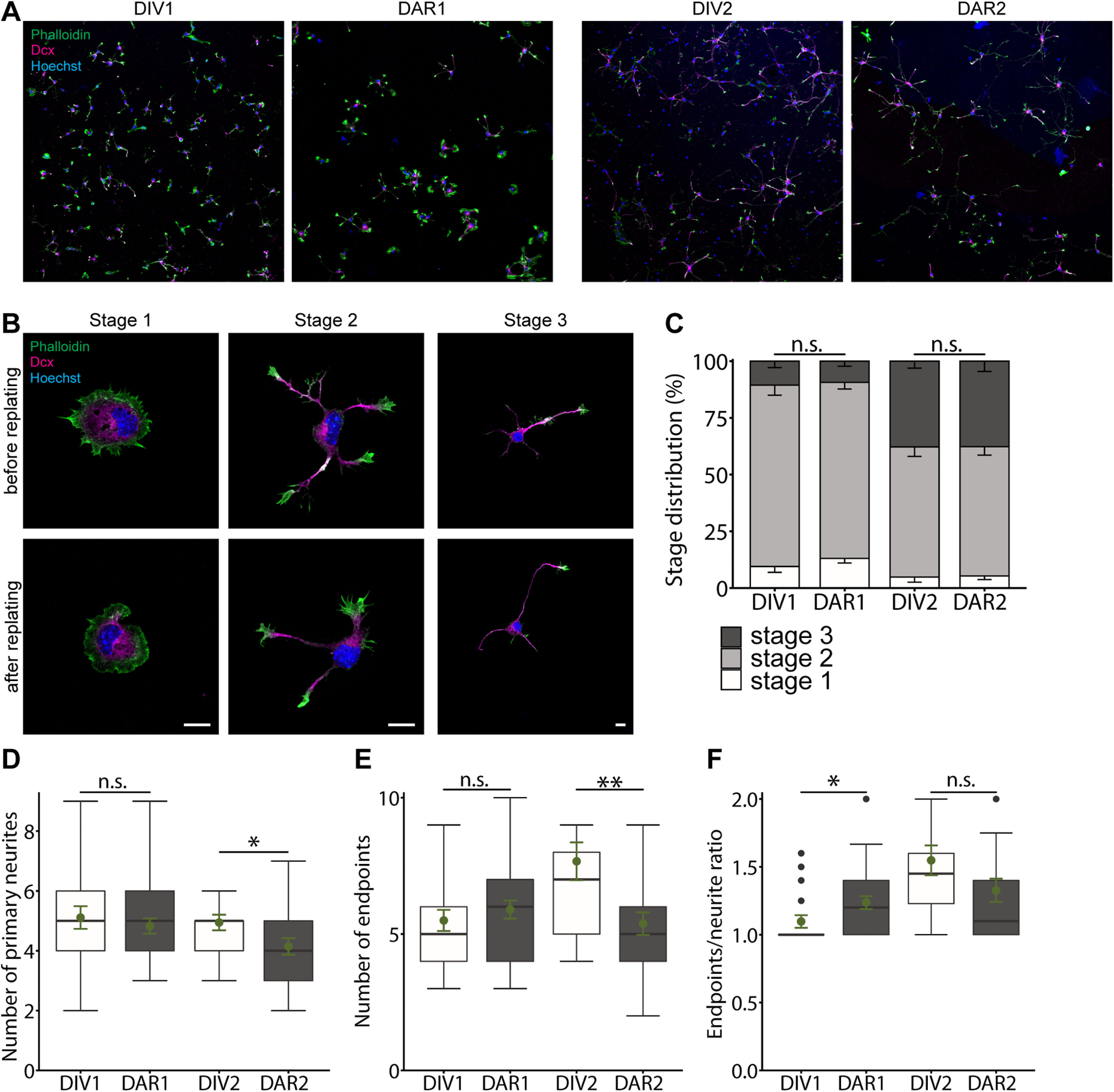
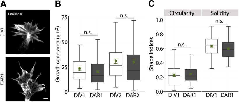
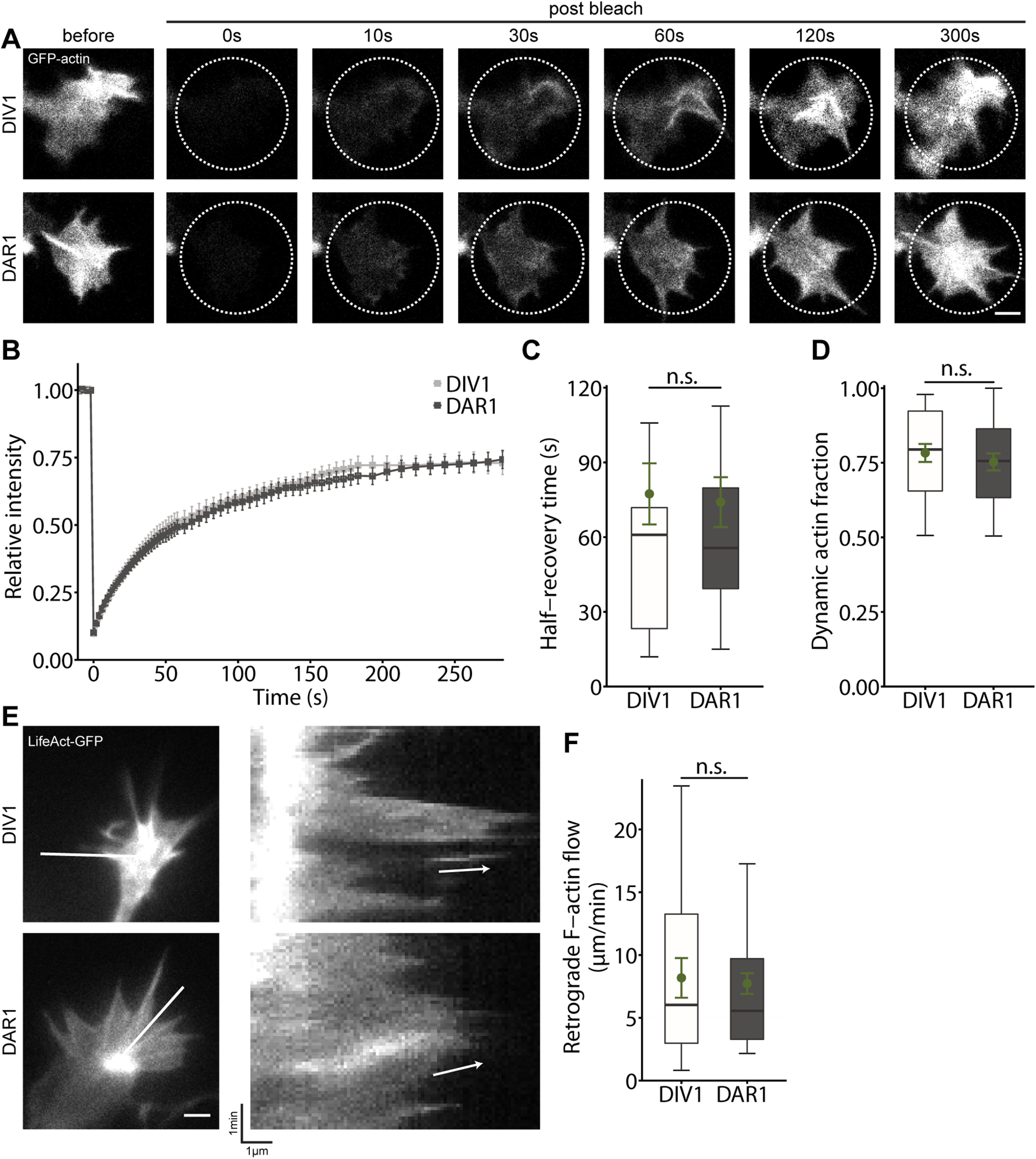
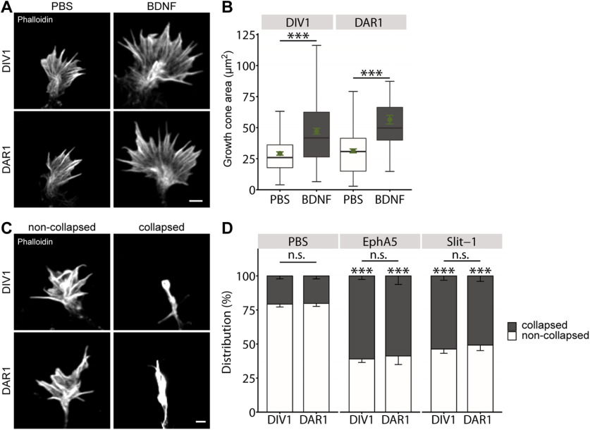
Neuron differentiation includes formation and outgrowth of neurites that differentiate into axons or dendrites. Directed neurite outgrowth is controlled by growth cones that protrude and retract actin-rich structures to sense environmental cues. These cues control local actin filament dynamics, steer growth cones toward attractants and away from repellents, and navigate neurites through the developing brain. Rodent hippocampal neurons are widely used to study the mechanisms underlying neuron differentiation. Genetic manipulation of isolated neurons including gene inactivation or reporter gene expression can be achieved by classical transfections methods, but these methods are restricted to neurons cultured for several days, after neurite formation or outgrowth. Instead, electroporation allows gene manipulation before seeding. However, reporter gene expression usually takes up to 24 h, and time course of gene inactivation depends on the half live of the targeted mRNA and gene product. Hence, these methods do not allow to study early aspects of neuron differentiation. In the present study, we provide a detailed protocol in which we combined electroporation-based gene manipulation of mouse hippocampal neurons before initial seeding with a replating step after 2 d (DIV) that resets neurons into an undifferentiated stage. By categorizing neurons according to their differentiation stage, thorough morphometric analyses, live imaging of actin dynamics in growth cones as well as guidance cue-mediated growth cone morphologic changes, we demonstrate that differentiation and function of replated neurons did not differ from non-replated neurons. In summary, we provide a protocol that allows to thoroughly characterize differentiation of mouse primary hippocampal neurons.
神经元分化包括神经突的形成和生长,神经突可分化为轴突或树突。定向神经突生长受生长锥控制,生长锥通过伸出和缩回富含肌动蛋白的结构来感知环境线索。这些线索控制局部肌动蛋白丝的动态变化,引导生长锥朝向吸引物并远离排斥物,并引导神经突在发育中的大脑中导航。啮齿动物海马神经元被广泛用于研究神经元分化的潜在机制。通过经典转染方法可以实现对分离神经元的基因操作,包括基因失活或报告基因表达,但这些方法仅限于在神经突形成或生长后培养数天的神经元。相反,电穿孔允许在接种前进行基因操作。然而,报告基因的表达通常需要长达24小时,基因失活的时间进程取决于靶向mRNA和基因产物的半衰期。因此,这些方法不允许研究神经元分化的早期阶段。在本研究中,我们提供了一个详细的方案,其中我们将初始接种前基于电穿孔的小鼠海马神经元基因操作与2天(DIV)后重新接种步骤相结合,该步骤将神经元重置为未分化阶段。通过根据神经元的分化阶段对其进行分类、进行全面的形态计量分析、对生长锥中肌动蛋白动力学进行实时成像以及对引导线索介导的生长锥形态变化进行分析,我们证明重新接种的神经元的分化和功能与未重新接种的神经元没有差异。总之,我们提供了一个方案,该方案允许全面表征小鼠原代海马神经元的分化。