Department of Science of Dental Materials, Bangladesh Dental College, Dhaka, Bangladesh.
Orthodontics Unit, School of Dental Sciences, Universiti Sains Malaysia, Kota Bharu, Kelantan, Malaysia.
Biomed Res Int. 2021 Apr 17;2021:6663683. doi: 10.1155/2021/6663683. eCollection 2021.
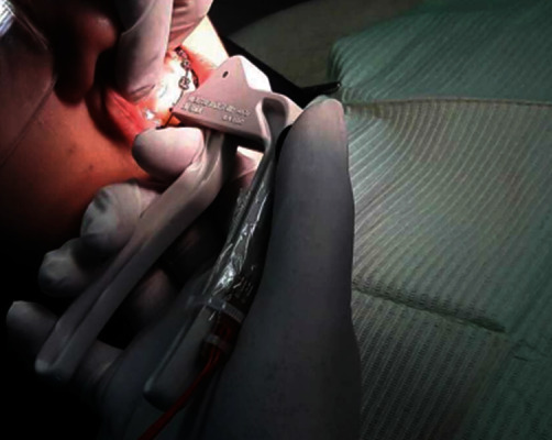
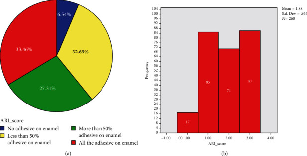
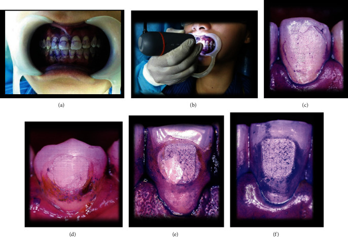
To compare the orthodontic bracket debonding force and assess the bracket failure pattern clinically between different teeth by a validated prototype debonding device. . Thirteen (13) patients at the end of comprehensive fixed orthodontic treatment, awaiting for bracket removal, were selected from the list. A total of 260 brackets from the central incisor to the second premolar in both jaws were debonded by a single clinician using a validated prototype debonding device equipped with a force sensitive resistor (FSR). Mean bracket debonding forces were specified to ten (10) groups of teeth. Following debonding, Intraoral microphotographs of the teeth were taken by the same clinician to assess the bracket failure pattern using a 4-point scale of adhesive remnant index (ARI). Statistical analysis included one-way ANOVA with post hoc Tukey HSD and independent sample -test to compare in vivo bracket debonding force, Cohen's kappa (), and a nonparametric Kruskal-Wallis test for the reliability and the assessment of ARI scoring.
A significant difference ( < 0.001) of mean debonding force was found between different types of teeth in vivo. Clinically, ARI scores were not significantly different ( = 0.921) between different groups, but overall higher scores were predominant.
Bracket debonding force should be measured on the same tooth from the same arch as the significant difference of mean debonding force exists between similar teeth of the upper and lower arches. The insignificant bracket failure pattern with higher ARI scores confirms less enamel damage irrespective of tooth types.
通过一种经过验证的原型脱轨装置比较不同牙齿的正畸托槽脱轨力,并评估托槽的脱轨模式。方法:从综合固定正畸治疗结束、等待托槽去除的患者名单中选择了 13 名患者。由同一位临床医生使用配备力敏电阻器(FSR)的经过验证的原型脱轨装置从上下颌的中切牙到第二前磨牙共脱轨 260 个托槽。指定十个(10)组牙齿的平均托槽脱轨力。脱轨后,同一位临床医生通过口腔内微摄影拍摄牙齿的照片,使用 4 分制的黏附残留指数(ARI)评估托槽的脱轨模式。统计分析包括单向方差分析(ANOVA),具有事后 Tukey HSD 和独立样本 t 检验,用于比较体内托槽脱轨力、Cohen's kappa(),以及可靠性的非参数 Kruskal-Wallis 检验和 ARI 评分的评估。结果:发现体内不同类型牙齿的平均脱轨力存在显著差异(<0.001)。临床上,不同组之间的 ARI 评分无显著差异(=0.921),但总体上较高的评分更为常见。结论:应该在上颌和下颌相同弓丝的相同牙齿上测量托槽脱轨力,因为上颌和下颌相似牙齿之间存在显著的平均脱轨力差异。较高 ARI 评分的托槽脱轨模式不明显,这证实了无论牙齿类型如何,釉质损伤较小。