Department of Cardiology, University Hospital of the RWTH Aachen, Aachen, Germany.
Zoological Institute, Department of Cell- and Neurobiology, Karlsruhe Institute of Technology (KIT), Karlsruhe, Germany.
Elife. 2021 May 11;10:e64020. doi: 10.7554/eLife.64020.
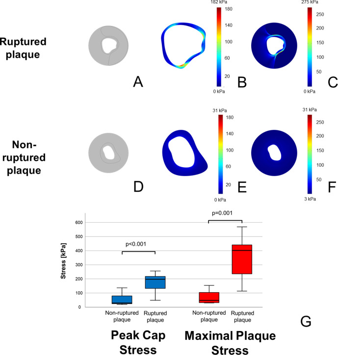
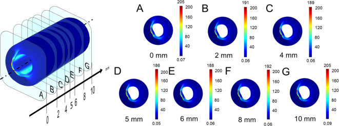
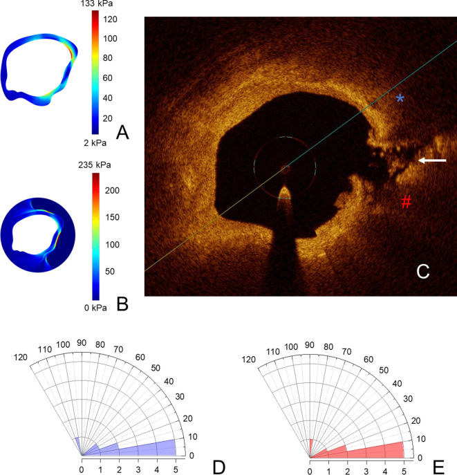
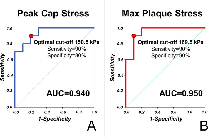
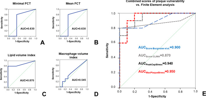
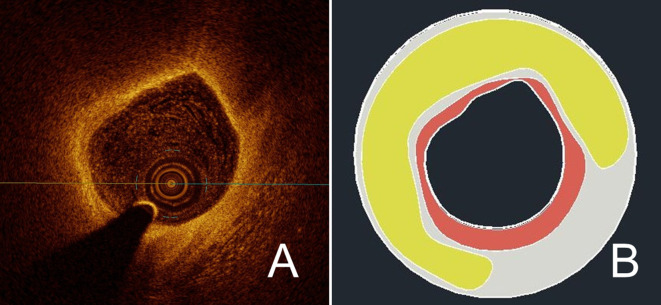
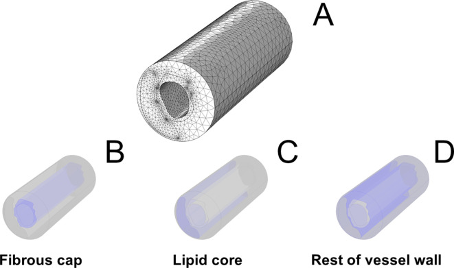
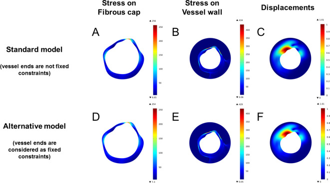
Plaque rupture occurs if stress within coronary lesions exceeds the protection exerted by the fibrous cap overlying the necrotic lipid core. However, very little is known about the biomechanical stress exerting this disrupting force. Employing optical coherence tomography (OCT), we generated plaque models and performed finite-element analysis to simulate stress distributions within the vessel wall in 10 ruptured and 10 non-ruptured lesions. In ruptured lesions, maximal stress within fibrous cap (peak cap stress [PCS]: 174 ± 67 vs. 52 ± 42 kPa, p<0.001) and vessel wall (maximal plaque stress [MPS]: 399 ± 233 vs. 90 ± 95 kPa, p=0.001) were significantly higher compared to non-ruptured plaques. Ruptures arose in the immediate proximity of maximal stress concentrations (angular distances: 21.8 ± 30.3° for PCS vs. 20.7 ± 23.7° for MPS); stress concentrations excellently predicted plaque rupture (area under the curve: 0.940 for PCS, 0.950 for MPS). This prediction of plaque rupture was superior to established vulnerability features such as fibrous cap thickness or macrophage infiltration. In conclusion, OCT-based finite-element analysis effectively assesses plaque biomechanics, which in turn predicts plaque rupture in patients. This highlights the importance of morpho-mechanic analysis assessing the disrupting effects of plaque stress.
斑块破裂发生于冠状动脉病变处的内应力超过覆盖在坏死脂质核心之上的纤维帽所能承受的保护力时。然而,人们对于这种破坏力量的生物力学压力知之甚少。我们采用光学相干断层扫描(OCT)生成斑块模型,并进行有限元分析,以模拟 10 个破裂和 10 个未破裂病变中血管壁内的应力分布。在破裂的病变中,纤维帽内的最大应力(峰值帽应力[PCS]:174 ± 67 与 52 ± 42 kPa,p<0.001)和血管壁内的最大斑块应力(MPS:399 ± 233 与 90 ± 95 kPa,p=0.001)明显高于未破裂的斑块。破裂发生在最大应力集中的附近(PCS 的角距离:21.8 ± 30.3°,MPS 的角距离:20.7 ± 23.7°);应力集中可极好地预测斑块破裂(PCS 的曲线下面积:0.940,MPS 的曲线下面积:0.950)。这种对斑块破裂的预测优于纤维帽厚度或巨噬细胞浸润等已确立的易损性特征。总之,基于 OCT 的有限元分析可有效评估斑块生物力学,进而预测患者的斑块破裂。这凸显了评估斑块应力破坏作用的形态力学分析的重要性。