• 文献检索
  • 文档翻译
  • 深度研究
  • 学术资讯
  • Suppr Zotero 插件Zotero 插件
  • 邀请有礼
  • 套餐&价格
  • 历史记录
应用&插件
Suppr Zotero 插件Zotero 插件浏览器插件Mac 客户端Windows 客户端微信小程序
定价
高级版会员购买积分包购买API积分包
服务
文献检索文档翻译深度研究API 文档MCP 服务
关于我们
关于 Suppr公司介绍联系我们用户协议隐私条款
关注我们

Suppr 超能文献

核心技术专利:CN118964589B侵权必究
粤ICP备2023148730 号-1Suppr @ 2026

文献检索

告别复杂PubMed语法,用中文像聊天一样搜索,搜遍4000万医学文献。AI智能推荐,让科研检索更轻松。

立即免费搜索

文件翻译

保留排版,准确专业,支持PDF/Word/PPT等文件格式,支持 12+语言互译。

免费翻译文档

深度研究

AI帮你快速写综述,25分钟生成高质量综述,智能提取关键信息,辅助科研写作。

立即免费体验

固有钙化角:2 型糖尿病患者易损性冠状动脉斑块的一个新特征:一项光学相干断层成像研究。

Intrinsic calcification angle: a novel feature of the vulnerable coronary plaque in patients with type 2 diabetes: an optical coherence tomography study.

机构信息

Department of Cardiology, Medical Clinic I, University Hospital of the RWTH Aachen, Pauwelsstr. 30, 52074, Aachen, Germany.

Zoological Institute, Department of Cell- and Neurobiology, Karlsruhe Institute of Technology (KIT), Karlsruhe, Germany.

出版信息

Cardiovasc Diabetol. 2019 Sep 24;18(1):122. doi: 10.1186/s12933-019-0926-x.

DOI:10.1186/s12933-019-0926-x
PMID:31551093
原文链接:https://pmc.ncbi.nlm.nih.gov/articles/PMC6760065/
Abstract

BACKGROUND

Coronary calcification is associated with high risk for cardiovascular events. However, its impact on plaque vulnerability is incompletely understood. In the present study we defined the intrinsic calcification angle (ICA) as the angle externally projected by a vascular calcification and analyzed its role as novel feature of coronary plaque vulnerability in patients with type 2 diabetes.

METHODS

Optical coherence tomography was used to determine ICA in 219 calcifications from 56 patients with stable coronary artery disease (CAD) and 143 calcifications from 36 patients with acute coronary syndrome (ACS). We then used finite elements analysis to gain mechanistic insight into the effects of ICA.

RESULTS

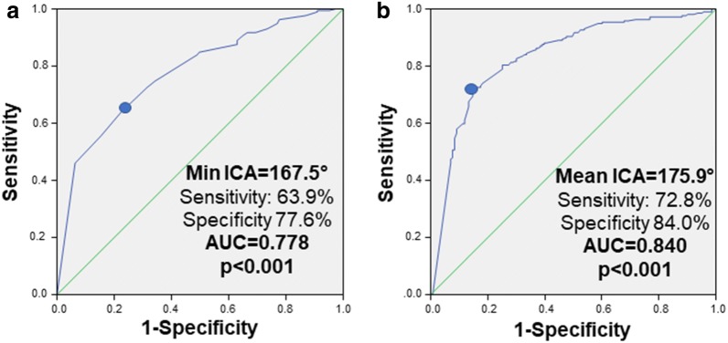
Minimal (139.8 ± 32.8° vs. 165.6 ± 21.6°, p < 0.001) and mean ICA (164.1 ± 14.3° vs. 176.0 ± 8.4°, p < 0.001) were lower in ACS vs. stable CAD patients. Mean ICA predicted ACS with very good diagnostic efficiency (AUC = 0.840, 95% CI 0.797-0.882, p < 0.001, optimal cut-off 175.9°); younger age (OR 0.95 per year, 95% CI 0.92-0.98, p = 0.002), male sex (OR 2.18, 95% CI 1.41-3.38, p < 0.001), lower HDL-cholesterol (OR 0.82 per 10 mg/dl, 95% CI 0.68-0.98, p = 0.029) and ACS (OR 14.71, 95% CI 8.47-25.64, p < 0.001) were determinants of ICA < 175.9°. A lower ICA predicted ACS (OR for 10°-variation 0.25, 95% CI 0.13-0.52, p < 0.001) independently from fibrous cap thickness, presence of macrophages or extension of lipid core. In finite elements analysis we confirmed that lower ICA causes increased stress on a lesion's fibrous cap; this effect was potentiated in more superficial calcifications and adds to the destabilizing role of smaller calcifications.

CONCLUSION

Our clinical and mechanistic data for the first time identify ICA as a novel feature of coronary plaque vulnerability.

摘要

背景

冠状动脉钙化与心血管事件的高风险相关。然而,其对斑块易损性的影响尚不完全清楚。在本研究中,我们将血管钙化的外部投影角度定义为固有钙化角(ICA),并分析了其在 2 型糖尿病患者中作为冠状动脉斑块易损性新特征的作用。

方法

使用光学相干断层扫描(OCT)确定 56 例稳定型冠状动脉疾病(CAD)患者的 219 个钙化灶和 36 例急性冠状动脉综合征(ACS)患者的 143 个钙化灶的 ICA。然后,我们使用有限元分析来深入了解 ICA 的影响。

结果

ACS 患者的最小 ICA(139.8±32.8° vs. 165.6±21.6°,p<0.001)和平均 ICA(164.1±14.3° vs. 176.0±8.4°,p<0.001)均低于稳定 CAD 患者。平均 ICA 对 ACS 具有非常好的诊断效率(AUC=0.840,95%CI 0.797-0.882,p<0.001,最佳截断值 175.9°);年龄较小(每年 0.95,95%CI 0.92-0.98,p=0.002)、男性(OR 2.18,95%CI 1.41-3.38,p<0.001)、较低的高密度脂蛋白胆固醇(每 10mg/dl 0.82,95%CI 0.68-0.98,p=0.029)和 ACS(OR 14.71,95%CI 8.47-25.64,p<0.001)是 ICA<175.9°的决定因素。较低的 ICA 预测 ACS(每 10°变化的 OR 0.25,95%CI 0.13-0.52,p<0.001)独立于纤维帽厚度、巨噬细胞存在或脂质核心延伸。在有限元分析中,我们证实较低的 ICA 会导致病变纤维帽上的应力增加;这种效应在更浅层的钙化中更为明显,并增加了较小钙化的不稳定性作用。

结论

我们的临床和机制数据首次将 ICA 确定为冠状动脉斑块易损性的一个新特征。

https://cdn.ncbi.nlm.nih.gov/pmc/blobs/c090/6760065/ff3a3a38f1cb/12933_2019_926_Fig4_HTML.jpg
https://cdn.ncbi.nlm.nih.gov/pmc/blobs/c090/6760065/27111af813ae/12933_2019_926_Fig1_HTML.jpg
https://cdn.ncbi.nlm.nih.gov/pmc/blobs/c090/6760065/3e5d6ca7f36b/12933_2019_926_Fig3_HTML.jpg
https://cdn.ncbi.nlm.nih.gov/pmc/blobs/c090/6760065/ff3a3a38f1cb/12933_2019_926_Fig4_HTML.jpg
https://cdn.ncbi.nlm.nih.gov/pmc/blobs/c090/6760065/27111af813ae/12933_2019_926_Fig1_HTML.jpg
https://cdn.ncbi.nlm.nih.gov/pmc/blobs/c090/6760065/3e5d6ca7f36b/12933_2019_926_Fig3_HTML.jpg
https://cdn.ncbi.nlm.nih.gov/pmc/blobs/c090/6760065/ff3a3a38f1cb/12933_2019_926_Fig4_HTML.jpg

相似文献

1
Intrinsic calcification angle: a novel feature of the vulnerable coronary plaque in patients with type 2 diabetes: an optical coherence tomography study.固有钙化角:2 型糖尿病患者易损性冠状动脉斑块的一个新特征:一项光学相干断层成像研究。
Cardiovasc Diabetol. 2019 Sep 24;18(1):122. doi: 10.1186/s12933-019-0926-x.
2
Type 2 diabetes mellitus is associated with a lower fibrous cap thickness but has no impact on calcification morphology: an intracoronary optical coherence tomography study.2 型糖尿病与较薄的纤维帽厚度相关,但对钙化形态无影响:一项冠状动脉光学相干断层成像研究。
Cardiovasc Diabetol. 2017 Dec 1;16(1):152. doi: 10.1186/s12933-017-0635-2.
3
Association Between Insulin Resistance and Coronary Plaque Vulnerability in Patients With Acute Coronary Syndromes: Insights From Optical Coherence Tomography.急性冠脉综合征患者胰岛素抵抗与冠状动脉斑块易损性之间的关联:光学相干断层扫描的见解
Angiology. 2019 Jul;70(6):539-546. doi: 10.1177/0003319718809931. Epub 2018 Nov 1.
4
Sex Differences in Nonculprit Coronary Plaque Microstructures on Frequency-Domain Optical Coherence Tomography in Acute Coronary Syndromes and Stable Coronary Artery Disease.急性冠状动脉综合征和稳定型冠状动脉疾病中基于频域光学相干断层扫描的非罪犯冠状动脉斑块微结构的性别差异
Circ Cardiovasc Imaging. 2016 Aug;9(8). doi: 10.1161/CIRCIMAGING.116.004506.
5
Prevalence, Predictors, and Clinical Presentation of a Calcified Nodule as Assessed by Optical Coherence Tomography.光学相干断层扫描评估的钙化结节的患病率、预测因素和临床表现。
JACC Cardiovasc Imaging. 2017 Aug;10(8):883-891. doi: 10.1016/j.jcmg.2017.05.013.
6
New insights into spotty calcification and plaque rupture in acute coronary syndrome: an optical coherence tomography study.急性冠状动脉综合征中斑点状钙化和斑块破裂的新见解:一项光学相干断层扫描研究
Heart Vessels. 2016 Dec;31(12):1915-1922. doi: 10.1007/s00380-016-0820-3. Epub 2016 Mar 5.
7
Plaque Morphology as Predictor of Late Plaque Events in Patients With Asymptomatic Type 2 Diabetes: A Long-Term Observational Study.斑块形态作为无症状 2 型糖尿病患者晚期斑块事件的预测因子:一项长期观察研究。
JACC Cardiovasc Imaging. 2019 Jul;12(7 Pt 2):1353-1363. doi: 10.1016/j.jcmg.2018.02.025. Epub 2018 May 16.
8
Calcified Plaques in Patients With Acute Coronary Syndromes.急性冠状动脉综合征患者的钙化斑块。
JACC Cardiovasc Interv. 2019 Mar 25;12(6):531-540. doi: 10.1016/j.jcin.2018.12.013.
9
Association between cholesterol crystals and culprit lesion vulnerability in patients with acute coronary syndrome: An optical coherence tomography study.急性冠状动脉综合征患者胆固醇结晶与罪犯病变易损性的关联:一项光学相干断层扫描研究
Atherosclerosis. 2016 Apr;247:111-7. doi: 10.1016/j.atherosclerosis.2016.02.010. Epub 2016 Feb 8.
10
New Insights into the Association between Fibrinogen and Coronary Atherosclerotic Plaque Vulnerability: An Intravascular Optical Coherence Tomography Study.纤维蛋白原与冠状动脉粥样硬化斑块易损性相关性的新认识:血管内光学相干断层成像研究。
Cardiovasc Ther. 2019 Apr 2;2019:8563717. doi: 10.1155/2019/8563717. eCollection 2019.

引用本文的文献

1
Percutaneous Revascularization of Thrombotic and Calcified Coronary Lesions.血栓性和钙化性冠状动脉病变的经皮血管重建术
J Clin Med. 2025 Jan 22;14(3):692. doi: 10.3390/jcm14030692.
2
In Silico Investigation of the Interlaminar and Mechanical Fracture of Arteries with Atheromatic Plaque during Angioplasty Treatment.血管成形术治疗期间动脉粥样硬化斑块动脉的层间和机械断裂的计算机模拟研究
Biomedicines. 2024 Sep 14;12(9):2105. doi: 10.3390/biomedicines12092105.
3
A Review Paper on Optical Coherence Tomography Evaluation of Coronary Calcification Pattern: Is It Relevant Today?

本文引用的文献

1
A transcriptomic model to predict increase in fibrous cap thickness in response to high-dose statin treatment: Validation by serial intracoronary OCT imaging.一种基于转录组学的模型,可预测大剂量他汀类药物治疗后纤维帽厚度的增加:通过连续冠状动脉 OCT 成像进行验证。
EBioMedicine. 2019 Jun;44:41-49. doi: 10.1016/j.ebiom.2019.05.007. Epub 2019 May 22.
2
Effect of Statin Therapy on Fibrous Cap Thickness in Coronary Plaque on Optical Coherence Tomography - Review and Meta-Analysis.他汀类药物治疗对光学相干断层扫描冠状动脉斑块纤维帽厚度的影响-综述和荟萃分析。
Circ J. 2019 Jun 25;83(7):1480-1488. doi: 10.1253/circj.CJ-18-1376. Epub 2019 May 22.
3
关于冠状动脉钙化模式的光学相干断层扫描评估的综述文章:如今它是否具有相关性?
J Cardiovasc Dev Dis. 2024 Jul 24;11(8):231. doi: 10.3390/jcdd11080231.
4
Association between triglyceride glucose index, coronary artery calcification and multivessel coronary disease in Chinese patients with acute coronary syndrome.甘油三酯-葡萄糖指数与冠状动脉钙化及多支血管病变在急性冠状动脉综合征中国患者中的相关性。
Cardiovasc Diabetol. 2022 Sep 16;21(1):187. doi: 10.1186/s12933-022-01615-4.
5
Lesion Geometry as Assessed by Optical Coherence Tomography Is Related to Myocardial Ischemia as Determined by Cardiac Magnetic Resonance Imaging.光学相干断层扫描评估的病变几何形状与心脏磁共振成像确定的心肌缺血相关。
J Clin Med. 2021 Jul 28;10(15):3342. doi: 10.3390/jcm10153342.
6
Coronary plaque composition influences biomechanical stress and predicts plaque rupture in a morpho-mechanic OCT analysis.基于形态力学光学相干断层成像分析的冠状动脉斑块成分对生物力学应激的影响及其与斑块破裂的相关性研究。
Elife. 2021 May 11;10:e64020. doi: 10.7554/eLife.64020.
7
Quantitative Flow Ratio Is Related to Intraluminal Coronary Stenosis Parameters as Assessed with Optical Coherence Tomography.定量血流比与光学相干断层扫描评估的冠状动脉管腔内狭窄参数相关。
J Clin Med. 2021 Apr 24;10(9):1856. doi: 10.3390/jcm10091856.
8
Reduced oxidized LDL in T2D plaques is associated with a greater statin usage but not with future cardiovascular events.在 T2D 斑块中,氧化型 LDL 减少与他汀类药物的使用量增加有关,但与未来的心血管事件无关。
Cardiovasc Diabetol. 2020 Dec 14;19(1):214. doi: 10.1186/s12933-020-01189-z.
9
Contemporary plaque imaging for risk stratification of coronary artery disease: Are we getting there?用于冠状动脉疾病风险分层的当代斑块成像:我们正在实现目标吗?
Int J Cardiol Heart Vasc. 2020 Nov 24;31:100678. doi: 10.1016/j.ijcha.2020.100678. eCollection 2020 Dec.
10
Prognostic irrelevance of plaque vulnerability following plaque sealing in high-risk patients with type 2 diabetes: an optical coherence tomography study.斑块易损性在斑块封闭后对 2 型糖尿病高危患者预后无影响:光学相干断层扫描研究。
Cardiovasc Diabetol. 2020 Nov 12;19(1):192. doi: 10.1186/s12933-020-01168-4.
Co-localization of plaque macrophages with calcification is associated with a more vulnerable plaque phenotype and a greater calcification burden in coronary target segments as determined by OCT.
光学相干断层扫描(OCT)显示,斑块巨噬细胞与钙化的共存与更易损斑块表型以及冠状动脉靶段更大的钙化负担相关。
PLoS One. 2018 Oct 24;13(10):e0205984. doi: 10.1371/journal.pone.0205984. eCollection 2018.
4
Coronary Plaque Characteristics in Patients With Diabetes Mellitus Who Presented With Acute Coronary Syndromes.糖尿病患者急性冠状动脉综合征的冠状动脉斑块特征。
J Am Heart Assoc. 2018 Jul 13;7(14):e009245. doi: 10.1161/JAHA.118.009245.
5
Non-culprit plaque characteristics in acute coronary syndrome patients with raised hemoglobinA1c: an intravascular optical coherence tomography study.糖化血红蛋白升高的急性冠脉综合征患者非罪犯斑块特征:血管内光学相干断层成像研究。
Cardiovasc Diabetol. 2018 Jun 15;17(1):90. doi: 10.1186/s12933-018-0729-5.
6
Predictors for target lesion microcalcifications in patients with stable coronary artery disease: an optical coherence tomography study.稳定型冠状动脉疾病患者靶病变微钙化的预测因素:一项光学相干断层成像研究。
Clin Res Cardiol. 2018 Sep;107(9):763-771. doi: 10.1007/s00392-018-1243-1. Epub 2018 Apr 13.
7
Type 2 diabetes mellitus is associated with a lower fibrous cap thickness but has no impact on calcification morphology: an intracoronary optical coherence tomography study.2 型糖尿病与较薄的纤维帽厚度相关,但对钙化形态无影响:一项冠状动脉光学相干断层成像研究。
Cardiovasc Diabetol. 2017 Dec 1;16(1):152. doi: 10.1186/s12933-017-0635-2.
8
Prevalence of Subclinical Coronary Artery Disease in Masters Endurance Athletes With a Low Atherosclerotic Risk Profile.低动脉粥样硬化风险特征的耐力项目大师运动员的亚临床冠状动脉疾病的流行率。
Circulation. 2017 Jul 11;136(2):126-137. doi: 10.1161/CIRCULATIONAHA.116.026964. Epub 2017 May 2.
9
Three-dimensional volumetric assessment of coronary artery calcification in patients with stable coronary artery disease by OCT.应用 OCT 对稳定性冠心病患者冠状动脉钙化进行三维容积评估。
EuroIntervention. 2017 Jun 20;13(3):312-319. doi: 10.4244/EIJ-D-16-00139.
10
Measures of Diagnostic Accuracy: Basic Definitions.诊断准确性的测量:基本定义。
EJIFCC. 2009 Jan 20;19(4):203-11. eCollection 2009 Jan.