Jinan University Institute of Tumor Pharmacology, College of Pharmacy, Jinan University, Guangzhou, China.
Integrated Chinese and Western Medicine Postdoctoral Research Station, Jinan University, Guangzhou, China.
Signal Transduct Target Ther. 2021 May 12;6(1):176. doi: 10.1038/s41392-021-00563-x.
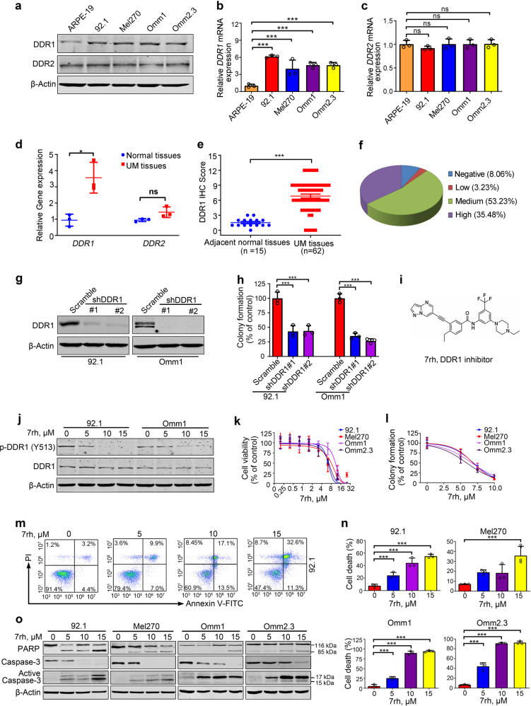
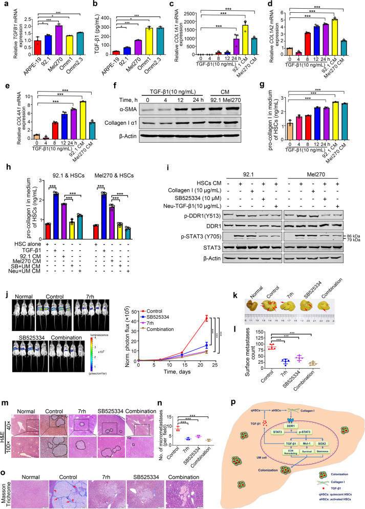
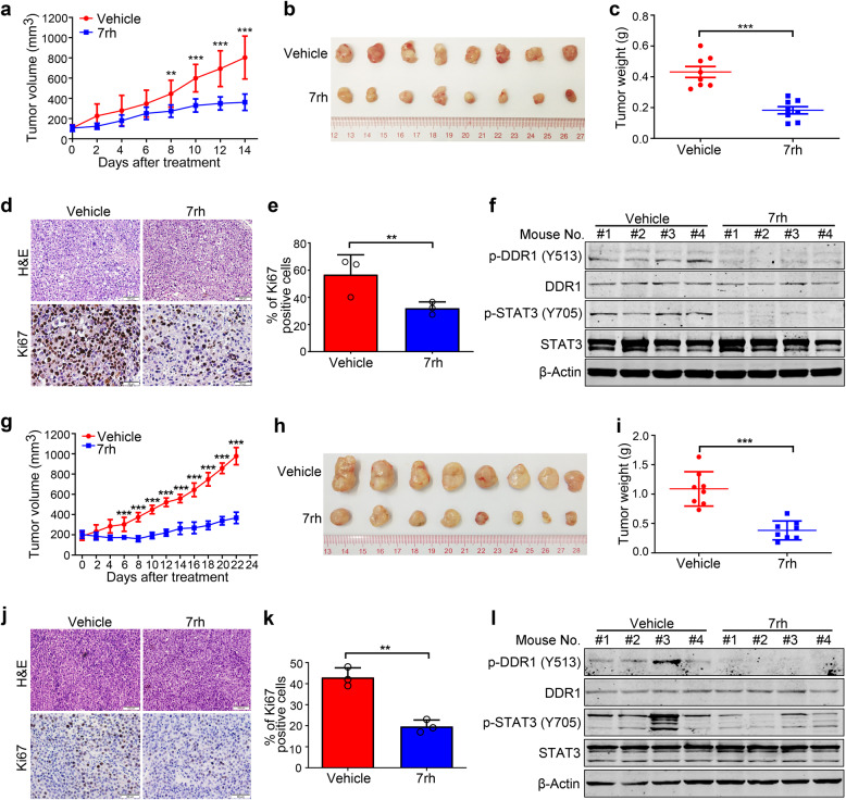
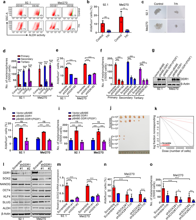
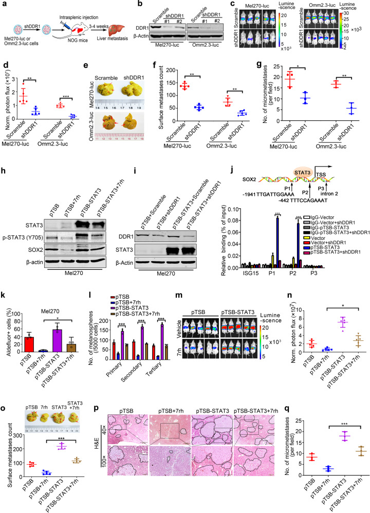
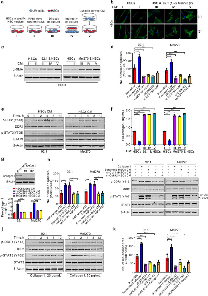
Colonization is believed a rate-limiting step of metastasis cascade. However, its underlying mechanism is not well understood. Uveal melanoma (UM), which is featured with single organ liver metastasis, may provide a simplified model for realizing the complicated colonization process. Because DDR1 was identified to be overexpressed in UM cell lines and specimens, and abundant pathological deposition of extracellular matrix collagen, a type of DDR1 ligand, was noted in the microenvironment of liver in metastatic patients with UM, we postulated the hypothesis that DDR1 and its ligand might ignite the interaction between UM cells and their surrounding niche of liver thereby conferring strengthened survival, proliferation, stemness and eventually promoting metastatic colonization in liver. We tested this hypothesis and found that DDR1 promoted these malignant cellular phenotypes and facilitated metastatic colonization of UM in liver. Mechanistically, UM cells secreted TGF-β1 which induced quiescent hepatic stellate cells (qHSCs) into activated HSCs (aHSCs) which secreted collagen type I. Such a remodeling of extracellular matrix, in turn, activated DDR1, strengthening survival through upregulating STAT3-dependent Mcl-1 expression, enhancing stemness via upregulating STAT3-dependent SOX2, and promoting clonogenicity in cancer cells. Targeting DDR1 by using 7rh, a specific inhibitor, repressed proliferation and survival in vitro and in vivo outgrowth. More importantly, targeting cancer cells by pharmacological inactivation of DDR1 or targeting microenvironmental TGF-β1-collagen I loop exhibited a prominent anti-metastasis effect in mice. In conclusion, targeting DDR1 signaling and TGF-β signaling may be a novel approach to diminish hepatic metastasis in UM.
定植被认为是转移级联的限速步骤。然而,其潜在机制尚不清楚。葡萄膜黑色素瘤(UM)以单一器官肝转移为特征,可能为实现复杂的定植过程提供了简化模型。因为 DDR1 在 UM 细胞系和标本中被鉴定为过度表达,并且在转移性 UM 患者的肝脏微环境中观察到丰富的细胞外基质胶原的病理性沉积,一种 DDR1 配体,我们假设假设 DDR1 及其配体可能引发 UM 细胞与其周围肝巢之间的相互作用,从而赋予更强的生存、增殖、干性,最终促进肝转移定植。我们检验了这个假设,发现 DDR1 促进了这些恶性细胞表型,并促进了 UM 在肝脏中的转移定植。从机制上讲,UM 细胞分泌 TGF-β1,诱导静止的肝星状细胞(qHSCs)转化为活化的肝星状细胞(aHSCs),后者分泌 I 型胶原。这种细胞外基质的重塑反过来激活了 DDR1,通过上调 STAT3 依赖性 Mcl-1 表达增强了细胞的生存能力,通过上调 STAT3 依赖性 SOX2 增强了干性,并促进了癌细胞的克隆形成能力。使用 7rh(一种特异性抑制剂)靶向 DDR1,抑制了体外和体内生长的增殖和存活。更重要的是,通过药理学失活 DDR1 靶向癌细胞或靶向微环境 TGF-β1-胶原 I 环在小鼠中表现出显著的抗转移作用。总之,靶向 DDR1 信号和 TGF-β 信号可能是减少 UM 肝转移的新方法。