Oi Marina, Maruhashi Takaaki, Kumazawa Kenichi, Iwakawa Saori, Kurihara Yutaro, Wato Jyunpei, Niimi Yuta, Takeda Akira, Asari Yasushi
Department of Emergency and Critical Care Medicine Kitasato University School of Medicine Sagamihara Japan.
Department of Plastic and Reconstructive Surgery Kitasato University Hospital Sagamihara Japan.
Acute Med Surg. 2021 May 3;8(1):e642. doi: 10.1002/ams2.642. eCollection 2021 Jan-Dec.
Skin and soft tissue infections are classified into cellulitis and necrotizing fasciitis, which are difficult to distinguish. Necrotizing fasciitis has a poor prognosis and requires immediate intensive care. The diagnostic gold standard is to incise the lesion to determine whether necrosis has reached the fascia. We aimed to show that these infections can be differentiated using near-infrared spectroscopy.
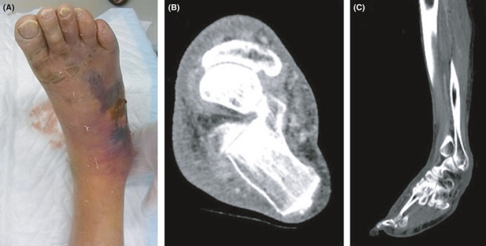
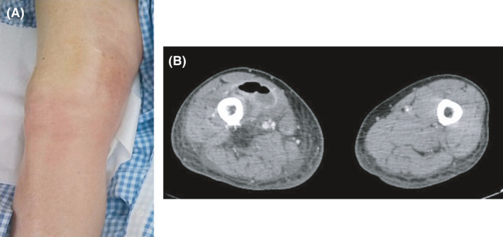
We describe two cases in an observational study about the utility of near-infrared spectroscopy. Case 1 involved a 77-year-old man with a chief complaint of pain, redness, and swelling in the right lower leg for 1 week. Computed tomography of his legs showed no gas formation. Case 2 involved an 82-year-old man. He visited another hospital because of pain, redness, and swelling in the right thigh. Based on the X-ray examination, necrotizing fasciitis was suspected, and he was transferred to our hospital.
In Case 1, the regional oxygen saturation value was lower on the lesion side (41%) than on the healthy side (55%). We confirmed the depth of invasion by incision, leading to a diagnosis of necrotizing fasciitis. In Case 2, the thigh's regional oxygen saturation was higher on the affected side (76%) than on the healthy side (61%). An incision was made for diagnosis, but the fascia was not necrotized. Thus, we diagnosed cellulitis and provided conservative treatment using antibiotics.
Near-infrared spectroscopy can be utilized to measure tissue blood flow, and it could be useful as a non-invasive diagnostic tool for skin and soft tissue infections.
皮肤和软组织感染分为蜂窝织炎和坏死性筋膜炎,二者难以区分。坏死性筋膜炎预后较差,需要立即进行重症监护。诊断金标准是切开病变部位以确定坏死是否累及筋膜。我们旨在表明可使用近红外光谱法区分这些感染。
我们在一项关于近红外光谱法效用的观察性研究中描述了两例病例。病例1为一名77岁男性,主要症状是右下肢疼痛、红肿1周。其腿部计算机断层扫描未显示气体形成。病例2为一名82岁男性。他因右大腿疼痛、红肿前往另一家医院就诊。基于X线检查,怀疑为坏死性筋膜炎,随后被转至我院。
病例1中,病变侧的局部氧饱和度值(41%)低于健康侧(55%)。我们通过切开确定了侵袭深度,诊断为坏死性筋膜炎。病例2中,患侧大腿的局部氧饱和度(76%)高于健康侧(61%)。为明确诊断进行了切开,但筋膜未坏死。因此,我们诊断为蜂窝织炎,并使用抗生素进行保守治疗。
近红外光谱法可用于测量组织血流,作为皮肤和软组织感染的非侵入性诊断工具可能有用。