• 文献检索
  • 文档翻译
  • 深度研究
  • 学术资讯
  • Suppr Zotero 插件Zotero 插件
  • 邀请有礼
  • 套餐&价格
  • 历史记录
应用&插件
Suppr Zotero 插件Zotero 插件浏览器插件Mac 客户端Windows 客户端微信小程序
定价
高级版会员购买积分包购买API积分包
服务
文献检索文档翻译深度研究API 文档MCP 服务
关于我们
关于 Suppr公司介绍联系我们用户协议隐私条款
关注我们

Suppr 超能文献

核心技术专利:CN118964589B侵权必究
粤ICP备2023148730 号-1Suppr @ 2026

文献检索

告别复杂PubMed语法,用中文像聊天一样搜索,搜遍4000万医学文献。AI智能推荐,让科研检索更轻松。

立即免费搜索

文件翻译

保留排版,准确专业,支持PDF/Word/PPT等文件格式,支持 12+语言互译。

免费翻译文档

深度研究

AI帮你快速写综述,25分钟生成高质量综述,智能提取关键信息,辅助科研写作。

立即免费体验

多光谱光栅扫描光声显微镜术利用结构和功能成像标记物鉴别特应性皮炎皮损和非皮损皮肤。

Multispectral raster-scanning optoacoustic mesoscopy differentiate lesional from non-lesional atopic dermatitis skin using structural and functional imaging markers.

作者信息

Li Xiuting, Moothanchery Mohesh, Kwa Cheng Yi, Tan Wan Ling, Yew Yik Weng, Thng Steven Tien Guan, Dinish U S, Attia Amalina Binte Ebrahim, Olivo Malini

机构信息

Translational Biophotonics Laboratory, Institute of Bioengineering and Bioimaging (IBB), Agency for Science, Technology and Research (A⁎STAR), Singapore.

National Skin Centre, Singapore.

出版信息

Photoacoustics. 2022 Aug 27;28:100399. doi: 10.1016/j.pacs.2022.100399. eCollection 2022 Dec.

DOI:10.1016/j.pacs.2022.100399
PMID:36090012
原文链接:https://pmc.ncbi.nlm.nih.gov/articles/PMC9450137/
Abstract

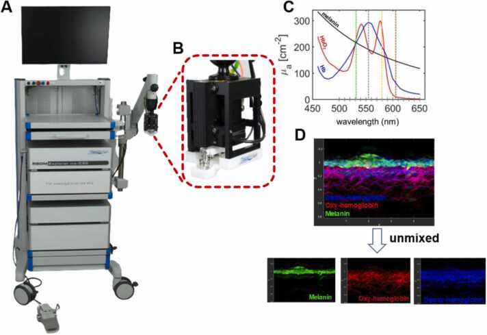
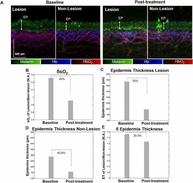
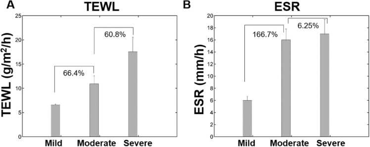
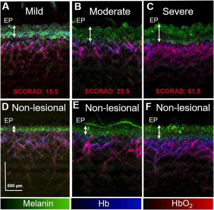
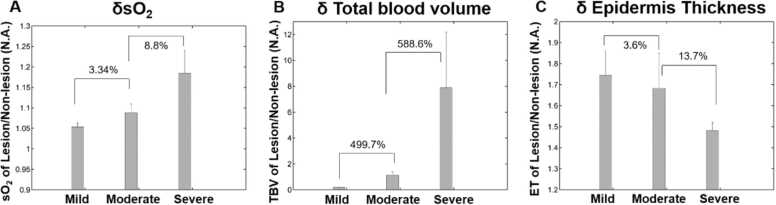
Atopic dermatitis (AD) is a chronic and pruritic skin inflammatory disease causing a significant burden to health care management and patient's quality of life. Seemingly healthy skin or non-lesional sites on AD patients still presents skin barrier defects and immune response, which can develop to AD at a later stage. To investigate further the balance between the epidermal barrier impairment and intrinsic immune dysregulation in AD, we exploited multispectral Raster-Scanning Optoacoustic Mesoscopy (ms-RSOM) to image lesional and non-lesional skin areas on AD patients of different severities non-invasively to elucidate their structural features and functional information. Herein, we demonstrate the objective assessment of AD severity using relative changes in oxygen saturation (δsO) levels in microvasculature along with other structural parameters such as relative changes in epidermis thickness (δET) and total blood volume (δTBV) between the lesional and non-lesional areas of the skin. We could observe an increasing trend for δsO and δTBV, which correlated well with the subjective clinical Scoring Atopic Dermatitis (SCORAD) for evaluating the severity. Notably, δET showed a decreasing trend with AD severity, indicating that the difference in epidermal thickness between lesional and non-lesional area of the skin decreases with AD severity. Our results also correlated well with conventional metrics such as trans-epidermal water loss (TEWL) and erythrosine sedimentation rate (ESR). We quantified the δsO and δET changes to objectively evaluate the treatment response before and four months after treatment using topical steroids and cyclosporine in one severe AD patient. We observed reduced δsO and δET post treatment. We envision that in future, functional and structural imaging metrics derived from ms-RSOM can be translated as objective markers to assess and stratify the severity of AD and understand the function of skin barrier dysfunctions and immune dysregulation. It could also be employed to monitor the treatment response of AD in regular clinical settings.

摘要

特应性皮炎(AD)是一种慢性瘙痒性皮肤炎症性疾病,给医疗管理和患者生活质量带来了沉重负担。AD患者看似健康的皮肤或非皮损部位仍存在皮肤屏障缺陷和免疫反应,这些情况在后期可能发展为AD。为了进一步研究AD中表皮屏障损伤与内在免疫失调之间的平衡,我们利用多光谱光栅扫描光声显微镜(ms-RSOM)对不同严重程度的AD患者的皮损和非皮损皮肤区域进行无创成像,以阐明其结构特征和功能信息。在此,我们通过微血管中氧饱和度(δsO)水平的相对变化以及其他结构参数,如皮肤皮损和非皮损区域之间表皮厚度(δET)和总血容量(δTBV)的相对变化,来展示对AD严重程度的客观评估。我们可以观察到δsO和δTBV呈上升趋势,这与用于评估严重程度的主观临床特应性皮炎评分(SCORAD)密切相关。值得注意的是,δET随着AD严重程度呈下降趋势,表明皮肤皮损和非皮损区域之间的表皮厚度差异随AD严重程度而减小。我们的结果也与传统指标如经表皮水分流失(TEWL)和红细胞沉降率(ESR)密切相关。我们对一名重度AD患者在使用外用类固醇和环孢素治疗前及治疗四个月后,对δsO和δET变化进行了量化,以客观评估治疗反应。我们观察到治疗后δsO和δET降低。我们设想,未来源自ms-RSOM的功能和结构成像指标可转化为客观标志物,用于评估和分层AD的严重程度,并了解皮肤屏障功能障碍和免疫失调的功能。它还可用于在常规临床环境中监测AD的治疗反应。

https://cdn.ncbi.nlm.nih.gov/pmc/blobs/91b0/9450137/114e42cd264a/gr5.jpg
https://cdn.ncbi.nlm.nih.gov/pmc/blobs/91b0/9450137/d37b1efed46f/gr1.jpg
https://cdn.ncbi.nlm.nih.gov/pmc/blobs/91b0/9450137/ae35377151e1/gr2.jpg
https://cdn.ncbi.nlm.nih.gov/pmc/blobs/91b0/9450137/939f79f2b37c/gr3.jpg
https://cdn.ncbi.nlm.nih.gov/pmc/blobs/91b0/9450137/28f11a43b950/gr4.jpg
https://cdn.ncbi.nlm.nih.gov/pmc/blobs/91b0/9450137/114e42cd264a/gr5.jpg

相似文献

1
Multispectral raster-scanning optoacoustic mesoscopy differentiate lesional from non-lesional atopic dermatitis skin using structural and functional imaging markers.多光谱光栅扫描光声显微镜术利用结构和功能成像标记物鉴别特应性皮炎皮损和非皮损皮肤。
Photoacoustics. 2022 Aug 27;28:100399. doi: 10.1016/j.pacs.2022.100399. eCollection 2022 Dec.
2
Structural and functional imaging of psoriasis for severity assessment and quantitative monitoring of treatment response using high-resolution optoacoustic imaging.利用高分辨率光声成像对银屑病进行结构和功能成像,以评估严重程度并定量监测治疗反应。
Photoacoustics. 2024 Apr 23;38:100611. doi: 10.1016/j.pacs.2024.100611. eCollection 2024 Aug.
3
Raster-scanning optoacoustic mesoscopy biomarkers for atopic dermatitis skin lesions.用于特应性皮炎皮肤病变的光栅扫描光声显微镜生物标志物
Photoacoustics. 2023 May 25;31:100513. doi: 10.1016/j.pacs.2023.100513. eCollection 2023 Jun.
4
Altered Levels of Sphingosine, Sphinganine and Their Ceramides in Atopic Dermatitis Are Related to Skin Barrier Function, Disease Severity and Local Cytokine Milieu.特应性皮炎中神经酰胺、鞘氨醇和它们的水平改变与皮肤屏障功能、疾病严重程度和局部细胞因子环境有关。
Int J Mol Sci. 2020 Mar 13;21(6):1958. doi: 10.3390/ijms21061958.
5
Pyrrolidone carboxylic acid levels or caspase-14 expression in the corneocytes of lesional skin correlates with clinical severity, skin barrier function and lesional inflammation in atopic dermatitis.病变皮肤角质形成细胞中的吡咯烷酮羧酸水平或半胱天冬酶-14表达与特应性皮炎的临床严重程度、皮肤屏障功能及病变炎症相关。
J Dermatol Sci. 2014 Dec;76(3):231-9. doi: 10.1016/j.jdermsci.2014.09.004. Epub 2014 Sep 30.
6
Potential of high-frequency ultrasonography in the management of atopic dermatitis.高频超声检查在特应性皮炎管理中的潜力。
Exp Ther Med. 2019 Feb;17(2):1073-1077. doi: 10.3892/etm.2018.6984. Epub 2018 Nov 16.
7
Model learning analysis of 3D optoacoustic mesoscopy images for the classification of atopic dermatitis.用于特应性皮炎分类的三维光声显微镜图像的模型学习分析
Biomed Opt Express. 2021 May 27;12(6):3671-3683. doi: 10.1364/BOE.415105. eCollection 2021 Jun 1.
8
Use of Tape Strips to Detect Immune and Barrier Abnormalities in the Skin of Children With Early-Onset Atopic Dermatitis.使用胶带条检测早发性特应性皮炎儿童皮肤的免疫和屏障异常。
JAMA Dermatol. 2019 Dec 1;155(12):1358-1370. doi: 10.1001/jamadermatol.2019.2983.
9
Microvascular imaging and monitoring of hemodynamic changes in the skin during arterial-venous occlusion using multispectral raster-scanning optoacoustic mesoscopy.使用多光谱光栅扫描光声显微镜对动静脉闭塞期间皮肤的微血管成像及血流动力学变化监测。
Photoacoustics. 2021 Apr 20;22:100268. doi: 10.1016/j.pacs.2021.100268. eCollection 2021 Jun.
10
Transient receptor potential vanilloid 3 expression is increased in non-lesional skin of atopic dermatitis patients.瞬时受体电位香草酸 3 表达在特应性皮炎患者非皮损皮肤中增加。
Exp Dermatol. 2022 May;31(5):807-813. doi: 10.1111/exd.14530. Epub 2022 Feb 4.

引用本文的文献

1
Three-Dimensional Curved Workflow-Based Optical Coherence Tomography Angiography for Enhancing Atopic Dermatitis Theranostics.基于三维曲线工作流程的光学相干断层扫描血管造影术用于增强特应性皮炎的诊疗。
Research (Wash D C). 2025 Aug 6;8:0778. doi: 10.34133/research.0778. eCollection 2025.
2
Pilot study on photoacoustic imaging and confocal Raman spectroscopy-derived biomarkers for assessing structural and physiological skin changes in atopic dermatitis and metabolic diseases.用于评估特应性皮炎和代谢性疾病中皮肤结构和生理变化的光声成像和共聚焦拉曼光谱衍生生物标志物的初步研究。
Biomed Opt Express. 2025 Jun 27;16(7):2986-3000. doi: 10.1364/BOE.559491. eCollection 2025 Jul 1.
3

本文引用的文献

1
Effects of Intermittent Treatment with Topical Corticosteroids and Calcineurin Inhibitors on Epidermal and Dermal Thickness Using Optical Coherence Tomography and Ultrasound.应用光学相干断层扫描和超声评估间断性外用皮质类固醇和钙调磷酸酶抑制剂对表皮和真皮厚度的影响。
Skin Pharmacol Physiol. 2022;35(1):41-50. doi: 10.1159/000518214. Epub 2021 Jul 8.
2
Model learning analysis of 3D optoacoustic mesoscopy images for the classification of atopic dermatitis.用于特应性皮炎分类的三维光声显微镜图像的模型学习分析
Biomed Opt Express. 2021 May 27;12(6):3671-3683. doi: 10.1364/BOE.415105. eCollection 2021 Jun 1.
3
Microvascular imaging and monitoring of hemodynamic changes in the skin during arterial-venous occlusion using multispectral raster-scanning optoacoustic mesoscopy.
3D photoacoustic and ultrasound analysis of hypopigmented skin lesions: A pilot study.
色素减退性皮肤病变的三维光声和超声分析:一项初步研究。
Photoacoustics. 2025 Mar 8;43:100705. doi: 10.1016/j.pacs.2025.100705. eCollection 2025 Jun.
4
Expert Consensus on Big Data Collection of Skin and Appendage Disease Phenotypes in Chinese.《中国皮肤及附属器疾病表型大数据采集专家共识》
Phenomics. 2024 Aug 19;4(3):269-292. doi: 10.1007/s43657-023-00142-w. eCollection 2024 Jun.
5
Structural and functional imaging of psoriasis for severity assessment and quantitative monitoring of treatment response using high-resolution optoacoustic imaging.利用高分辨率光声成像对银屑病进行结构和功能成像,以评估严重程度并定量监测治疗反应。
Photoacoustics. 2024 Apr 23;38:100611. doi: 10.1016/j.pacs.2024.100611. eCollection 2024 Aug.
6
Photoacoustic imaging for cutaneous melanoma assessment: a comprehensive review.光声成像在皮肤黑色素瘤评估中的应用:全面综述。
J Biomed Opt. 2024 Jan;29(Suppl 1):S11518. doi: 10.1117/1.JBO.29.S1.S11518. Epub 2024 Jan 12.
7
Functional photoacoustic imaging: from nano- and micro- to macro-scale.功能性光声成像:从纳米和微米尺度到宏观尺度
Nano Converg. 2023 Jun 19;10(1):29. doi: 10.1186/s40580-023-00377-3.
8
Raster-scanning optoacoustic mesoscopy biomarkers for atopic dermatitis skin lesions.用于特应性皮炎皮肤病变的光栅扫描光声显微镜生物标志物
Photoacoustics. 2023 May 25;31:100513. doi: 10.1016/j.pacs.2023.100513. eCollection 2023 Jun.
9
Transrectal Absorber Guide Raster-Scanning Optoacoustic Mesoscopy for Label-Free In Vivo Assessment of Colitis.经直肠吸收器引导光栅扫描光声介观术用于无标记体内评估结肠炎。
Adv Sci (Weinh). 2023 Jun;10(18):e2300564. doi: 10.1002/advs.202300564. Epub 2023 Apr 21.
10
quantitative photoacoustic monitoring of corticosteroid-induced vasoconstriction.定量光声监测皮质类固醇诱导的血管收缩。
J Biomed Opt. 2023 Aug;28(8):082805. doi: 10.1117/1.JBO.28.8.082805. Epub 2023 Feb 24.
使用多光谱光栅扫描光声显微镜对动静脉闭塞期间皮肤的微血管成像及血流动力学变化监测。
Photoacoustics. 2021 Apr 20;22:100268. doi: 10.1016/j.pacs.2021.100268. eCollection 2021 Jun.
4
Diagnosis of skin and soft tissue infections using near-infrared spectroscopy.使用近红外光谱法诊断皮肤和软组织感染
Acute Med Surg. 2021 May 3;8(1):e642. doi: 10.1002/ams2.642. eCollection 2021 Jan-Dec.
5
Photoacoustic imaging of hemodynamic changes in forearm skeletal muscle during cuff occlusion.袖带阻断期间前臂骨骼肌血流动力学变化的光声成像
Biomed Opt Express. 2020 Jul 23;11(8):4560-4570. doi: 10.1364/BOE.392221. eCollection 2020 Aug 1.
6
Raster-scanning optoacoustic mesoscopy imaging as an objective disease severity tool in atopic dermatitis patients.光栅扫描光声显微镜成像作为特应性皮炎患者客观疾病严重程度评估工具
J Am Acad Dermatol. 2021 Apr;84(4):1121-1123. doi: 10.1016/j.jaad.2020.06.045. Epub 2020 Jun 17.
7
Detecting hemodynamic changes in the foot vessels of diabetic patients by photoacoustic tomography.利用光声断层成像技术检测糖尿病患者足部血管的血液动力学变化。
J Biophotonics. 2020 Aug;13(8):e202000011. doi: 10.1002/jbio.202000011. Epub 2020 May 25.
8
Multispectral optoacoustic tomography of muscle perfusion and oxygenation under arterial and venous occlusion: A human pilot study.多光谱光声断层扫描在动脉和静脉阻塞下对肌肉灌注和氧合的研究:一项人体初步研究。
J Biophotonics. 2020 Jun;13(6):e201960169. doi: 10.1002/jbio.201960169. Epub 2020 Mar 25.
9
Investigation of morphological, vascular and biochemical changes in the skin of an atopic dermatitis (AD) patient in response to dupilumab using raster scanning optoacoustic mesoscopy (RSOM) and handheld confocal Raman spectroscopy (CRS).使用光栅扫描光声显微镜(RSOM)和手持式共聚焦拉曼光谱(CRS)研究特应性皮炎(AD)患者皮肤在接受度普利尤单抗治疗后的形态、血管和生化变化。
J Dermatol Sci. 2019 Sep;95(3):123-125. doi: 10.1016/j.jdermsci.2019.07.003. Epub 2019 Jul 12.
10
High-speed simultaneous multiscale photoacoustic microscopy.高速同步多尺度光声显微镜。
J Biomed Opt. 2019 Aug;24(8):1-7. doi: 10.1117/1.JBO.24.8.086001.