• 文献检索
  • 文档翻译
  • 深度研究
  • 学术资讯
  • Suppr Zotero 插件Zotero 插件
  • 邀请有礼
  • 套餐&价格
  • 历史记录
应用&插件
Suppr Zotero 插件Zotero 插件浏览器插件Mac 客户端Windows 客户端微信小程序
定价
高级版会员购买积分包购买API积分包
服务
文献检索文档翻译深度研究API 文档MCP 服务
关于我们
关于 Suppr公司介绍联系我们用户协议隐私条款
关注我们

Suppr 超能文献

核心技术专利:CN118964589B侵权必究
粤ICP备2023148730 号-1Suppr @ 2026

文献检索

告别复杂PubMed语法,用中文像聊天一样搜索,搜遍4000万医学文献。AI智能推荐,让科研检索更轻松。

立即免费搜索

文件翻译

保留排版,准确专业,支持PDF/Word/PPT等文件格式,支持 12+语言互译。

免费翻译文档

深度研究

AI帮你快速写综述,25分钟生成高质量综述,智能提取关键信息,辅助科研写作。

立即免费体验

中国原发性干燥综合征患者的角膜神经结构。

Corneal nerve structure in patients with primary Sjögren's syndrome in China.

机构信息

Department of Ophthalmology, Peking University People's Hospital, Eye Diseases and Optometry Institute, 11 Xizhimen South Street, Beijing, 100044, Xicheng District, China.

Beijing Key Laboratory of Diagnosis and Therapy of Retinal and Choroid Diseases, College of Optometry, Peking University Health Science Center, Beijing, China.

出版信息

BMC Ophthalmol. 2021 May 12;21(1):211. doi: 10.1186/s12886-021-01967-7.

DOI:10.1186/s12886-021-01967-7
PMID:33980205
原文链接:https://pmc.ncbi.nlm.nih.gov/articles/PMC8117565/
Abstract

BACKGROUND

The aim of this study was to evaluate the in vivo confocal microscopic morphology of corneal subbasal nerves and its relationship with clinical parameters in patients with primary Sjögren's syndrome in China.

METHODS

This was a case control study of 22 dry eye disease (DED) patients with primary Sjögren's syndrome (pSS) and 20 control subjects with non-Sjögren dry eye disease (NSDE). Each patient underwent an evaluation of ocular surface disease using the tear film break-up time (TBUT), noninvasive tear film break-up time (NIKBUT), noninvasive tear meniscus height (NIKTMH), corneal staining (National Eye Institute scale, NEI), Schirmer I test, meibography, and corneal subbasal nerve analysis with in vivo confocal microscopy (IVCM). The right eye of each subject was included in this study.

RESULTS

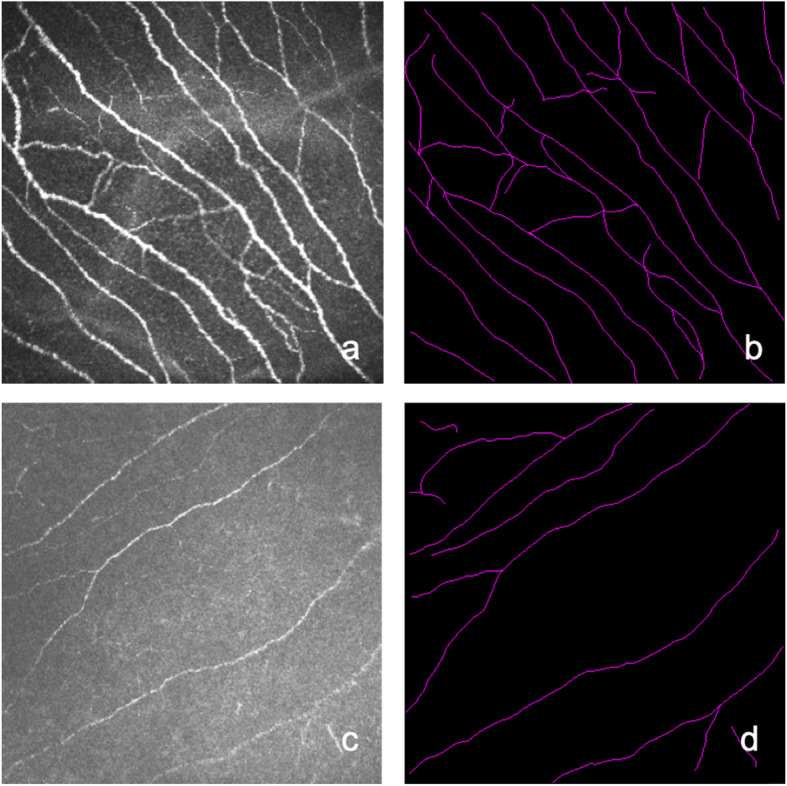
SS patients showed a shorter TBUT (P = 0.009) and Schirmer I test results (P = 0.028) than the NSDE group. However, there was no significant difference in NIKBUT between the two groups (P = 0.393). The nerve density of subbasal nerves, number of nerves and tortuosity of the SS group were significantly lower than those of the NSDE group (P = 0.001, P < 0.001 and P = 0.039, respectively). In the SS group, the mean nerve length was correlated with age and the Schirmer I test (r = - 0.519, P = 0.013 and r = 0.463, P = 0.035, respectively). Corneal staining was correlated with nerve density and the number of nerves (r = - 0.534, P = 0.013 and r = - 0.487, P = 0.025, respectively).

CONCLUSIONS

Sjögren syndrome dry eye (SSDE) patients have more severe clinical dry eye parameters than non-Sjögren dry eye disease (NSDE) patients. Compared with NSDE patients, we found that SSDE patients showed decreased corneal subbasal nerve density and numbers.

摘要

背景

本研究旨在评估中国原发性干燥综合征(pSS)患者角膜基底部神经的体内共聚焦显微镜形态及其与临床参数的关系。

方法

这是一项对 22 例干燥性眼病(DED)原发性干燥综合征(pSS)患者和 20 例非干燥性干燥综合征(NSDE)患者的病例对照研究。每位患者均接受了泪膜破裂时间(TBUT)、非侵入性泪膜破裂时间(NIKBUT)、非侵入性泪膜新月形高度(NIKTMH)、角膜染色(国家眼科研究所量表,NEI)、泪液分泌试验、泪膜造影和角膜基底部神经的评估分析采用体内共聚焦显微镜(IVCM)。每位患者的右眼均纳入本研究。

结果

SS 患者的 TBUT(P=0.009)和 Schirmer I 试验结果(P=0.028)均短于 NSDE 组。然而,两组之间的 NIKBUT 无显著差异(P=0.393)。SS 组的角膜基底部神经密度、神经数量和神经扭曲度均显著低于 NSDE 组(P=0.001、P<0.001 和 P=0.039)。在 SS 组中,平均神经长度与年龄和 Schirmer I 试验呈负相关(r=-0.519,P=0.013 和 r=-0.463,P=0.035)。角膜染色与神经密度和神经数量呈负相关(r=-0.534,P=0.013 和 r=-0.487,P=0.025)。

结论

干燥综合征(SS)干眼症(SSDE)患者的临床干眼症参数比非干燥综合征(NSDE)患者更严重。与 NSDE 患者相比,我们发现 SSDE 患者的角膜基底部神经密度和数量减少。

https://cdn.ncbi.nlm.nih.gov/pmc/blobs/1fe2/8117565/82946e206a6b/12886_2021_1967_Fig1_HTML.jpg

相似文献

1
Corneal nerve structure in patients with primary Sjögren's syndrome in China.中国原发性干燥综合征患者的角膜神经结构。
BMC Ophthalmol. 2021 May 12;21(1):211. doi: 10.1186/s12886-021-01967-7.
2
Correlation between corneal innervation and inflammation evaluated with confocal microscopy and symptomatology in patients with dry eye syndromes: a preliminary study.干眼综合征患者角膜神经支配与炎症的相关性:共聚焦显微镜检查及症状学评估的初步研究
Graefes Arch Clin Exp Ophthalmol. 2017 Sep;255(9):1771-1778. doi: 10.1007/s00417-017-3680-3. Epub 2017 May 20.
3
Corneal nerve structure and function in patients with non-sjogren dry eye: clinical correlations.非干燥综合征干眼患者的角膜神经结构和功能:临床相关性。
Invest Ophthalmol Vis Sci. 2013 Aug 1;54(8):5144-50. doi: 10.1167/iovs.13-12370.
4
Analysis of the first tear film break-up point in Sjögren's syndrome and non-Sjögren's syndrome dry eye patients.分析干燥综合征和非干燥综合征患者的第一次泪膜破裂点。
BMC Ophthalmol. 2022 Jan 3;22(1):1. doi: 10.1186/s12886-021-02233-6.
5
Tear Meniscus and Corneal Sub-basal Nerve Plexus Assessment in Primary Sjögren Syndrome and Sicca Syndrome Patients.原发性干燥综合征和干燥综合征患者的泪半月板和角膜基底神经丛评估
Cornea. 2019 Feb;38(2):221-228. doi: 10.1097/ICO.0000000000001800.
6
[Evaluation of ocular surface status and function in primary Sjögren's syndrome with hypothyroidism].[原发性干燥综合征合并甲状腺功能减退患者眼表状态及功能的评估]
Beijing Da Xue Xue Bao Yi Xue Ban. 2022 Aug 18;54(4):705-711. doi: 10.19723/j.issn.1671-167X.2022.04.020.
7
Evaluation of the corneal epithelium in non-Sjögren's and Sjögren's dry eyes: an in vivo confocal microscopy study using HRT III RCM.非干燥综合征和干燥综合征干眼患者角膜上皮的评估:一项使用HRT III RCM的活体共聚焦显微镜研究
BMC Ophthalmol. 2018 Dec 4;18(1):309. doi: 10.1186/s12886-018-0971-3.
8
Corneal subbasal nerve analysis in patients with primary Sjogren's syndrome: a novel objective grading method and clinical correlations.原发性干燥综合征患者的角膜基底膜下神经分析:一种新型客观分级方法及临床相关性
Int Ophthalmol. 2023 Mar;43(3):779-793. doi: 10.1007/s10792-022-02478-0. Epub 2022 Sep 2.
9
In vivo confocal microscopy findings of cornea and tongue mucosa in patients with Sjögren's syndrome.干燥综合征患者角膜和舌黏膜的共聚焦显微镜检查结果。
Arch Soc Esp Oftalmol (Engl Ed). 2024 Feb;99(2):56-61. doi: 10.1016/j.oftale.2023.11.016. Epub 2023 Dec 1.
10
Corneal confocal microscopy alterations in Sjögren's syndrome dry eye.干燥综合征干眼的角膜共焦显微镜检查改变
Acta Ophthalmol. 2017 Aug;95(5):e366-e372. doi: 10.1111/aos.13194. Epub 2016 Oct 24.

引用本文的文献

1
Geometrical Features of Subbasal Corneal Whorl-like Nerve Patterns in Dry Eye Disease: An In Vivo Confocal Microscopy Study.干眼疾病中角膜基底膜下漩涡状神经模式的几何特征:一项活体共聚焦显微镜研究
Ophthalmol Sci. 2024 Dec 5;5(2):100669. doi: 10.1016/j.xops.2024.100669. eCollection 2025 Mar-Apr.
2
CD4 T cells drive corneal nerve damage but not epitheliopathy in an acute aqueous-deficient dry eye model.CD4 T 细胞驱动角膜神经损伤,但不引起急性水性缺乏性干眼模型中的眼表疾病。
Proc Natl Acad Sci U S A. 2024 Nov 26;121(48):e2407648121. doi: 10.1073/pnas.2407648121. Epub 2024 Nov 19.
3
Segmentation and multiparametric evaluation of corneal whorl-like nerves for in vivo confocal microscopy images in dry eye disease.

本文引用的文献

1
Meibomian Gland Morphology: The Influence of Structural Variations on Gland Function and Ocular Surface Parameters.睑板腺形态:结构变异对腺体功能和眼表参数的影响。
Cornea. 2019 Dec;38(12):1506-1512. doi: 10.1097/ICO.0000000000002141.
2
Tear Meniscus and Corneal Sub-basal Nerve Plexus Assessment in Primary Sjögren Syndrome and Sicca Syndrome Patients.原发性干燥综合征和干燥综合征患者的泪半月板和角膜基底神经丛评估
Cornea. 2019 Feb;38(2):221-228. doi: 10.1097/ICO.0000000000001800.
3
Comparative Evaluation of Clinical Methods of Tear Film Stability Assessment: A Randomized Crossover Trial.
干眼病活体共聚焦显微镜图像中环状神经的分割和多参数评估。
BMJ Open Ophthalmol. 2024 Oct 7;9(1):e001861. doi: 10.1136/bmjophth-2024-001861.
4
Systematic review and meta-analysis of the diagnostic performance of lacrimal gland ultrasound elastography in primary Sjögren's syndrome.原发性干燥综合征患者泪腺超声弹性成像的诊断性能的系统评价和荟萃分析。
Int Ophthalmol. 2024 Jun 25;44(1):271. doi: 10.1007/s10792-024-03185-8.
5
Inflammation mechanism and anti-inflammatory therapy of dry eye.干眼的炎症机制与抗炎治疗
Front Med (Lausanne). 2024 Feb 14;11:1307682. doi: 10.3389/fmed.2024.1307682. eCollection 2024.
6
Rethinking Sjögren Beyond Inflammation: Considering the Role of Nerves in Driving Disease Manifestations.重新思考干燥综合征:关注神经在驱动疾病表现中的作用。
Eye Contact Lens. 2024 May 1;50(5):200-207. doi: 10.1097/ICL.0000000000001068. Epub 2024 Feb 7.
7
Sjögren's Versus Non-Sjögren's Ocular Features: Similar Symptoms, But Significantly Worse Signs.干燥综合征与非干燥综合征的眼部特征:相似的症状,但明显更严重的体征。
Invest Ophthalmol Vis Sci. 2024 Jan 2;65(1):23. doi: 10.1167/iovs.65.1.23.
8
The Autoimmune Rheumatic Disease Related Dry Eye and Its Association with Retinopathy.自身免疫性风湿病相关干眼及其与视网膜病变的关系。
Biomolecules. 2023 Apr 23;13(5):724. doi: 10.3390/biom13050724.
9
Corneal nerve loss and increased Langerhans cells are associated with disease severity in patients with rheumatoid arthritis.角膜神经丢失和朗格汉斯细胞增多与类风湿关节炎患者的疾病严重程度相关。
Eye (Lond). 2023 Oct;37(14):2950-2955. doi: 10.1038/s41433-023-02447-6. Epub 2023 Feb 18.
10
A synthetic tear protein resolves dry eye through promoting corneal nerve regeneration.一种人工泪液蛋白通过促进角膜神经再生来治疗干眼症。
Cell Rep. 2022 Aug 30;40(9):111307. doi: 10.1016/j.celrep.2022.111307.
比较评估泪膜稳定性评估的临床方法:一项随机交叉试验。
JAMA Ophthalmol. 2018 Mar 1;136(3):291-294. doi: 10.1001/jamaophthalmol.2017.6489.
4
TFOS DEWS II Report Executive Summary.TFOS DEWS II 报告执行摘要。
Ocul Surf. 2017 Oct;15(4):802-812. doi: 10.1016/j.jtos.2017.08.003. Epub 2017 Aug 8.
5
TFOS DEWS II pain and sensation report.TFOS DEWS II 干眼综合征相关眼部不适和眼表面感觉报告。
Ocul Surf. 2017 Jul;15(3):404-437. doi: 10.1016/j.jtos.2017.05.002. Epub 2017 Jul 20.
6
Minimising instilled volume reduces the impact of fluorescein on clinical measurements of tear film stability.减少滴入量可降低荧光素对泪膜稳定性临床测量的影响。
Cont Lens Anterior Eye. 2017 Jun;40(3):170-174. doi: 10.1016/j.clae.2017.01.004. Epub 2017 Feb 4.
7
Comparison of Standard Versus Wide-Field Composite Images of the Corneal Subbasal Layer by In Vivo Confocal Microscopy.体内共聚焦显微镜下角膜基底下层标准图像与宽视野复合图像的比较
Invest Ophthalmol Vis Sci. 2015 Sep;56(10):5801-7. doi: 10.1167/iovs.15-17434.
8
Effects of corneal nerve density on the response to treatment in dry eye disease.角膜神经密度对干眼疾病治疗反应的影响。
Ophthalmology. 2015 Apr;122(4):662-8. doi: 10.1016/j.ophtha.2014.11.006. Epub 2014 Dec 24.
9
In vivo confocal scanning laser microscopy in patients with primary Sjögren's syndrome: A monocentric experience.原发性干燥综合征患者的体内共聚焦扫描激光显微镜检查:单中心经验。
Mod Rheumatol. 2015 Jul;25(4):585-9. doi: 10.3109/14397595.2014.979523. Epub 2014 Dec 15.
10
Mosaicking the subbasal nerve plexus by guided eye movements.通过引导性眼球运动对基底神经丛进行拼接。
Invest Ophthalmol Vis Sci. 2014 Aug 26;55(9):6082-9. doi: 10.1167/iovs.14-14698.