Madhavan Ajay A, Eckel Laurence J, Carr Carrie M, Diehn Felix E, Lehman Vance T
Division of Neuroradiology, Department of Radiology, Mayo Clinic, 200 First St SW, Rochester, Minnesota 55905.
Radiol Case Rep. 2021 Apr 16;16(6):1499-1503. doi: 10.1016/j.radcr.2021.03.025. eCollection 2021 Jun.
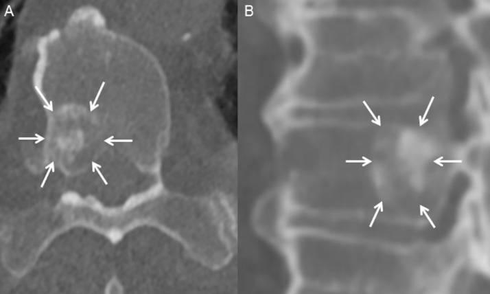
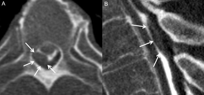
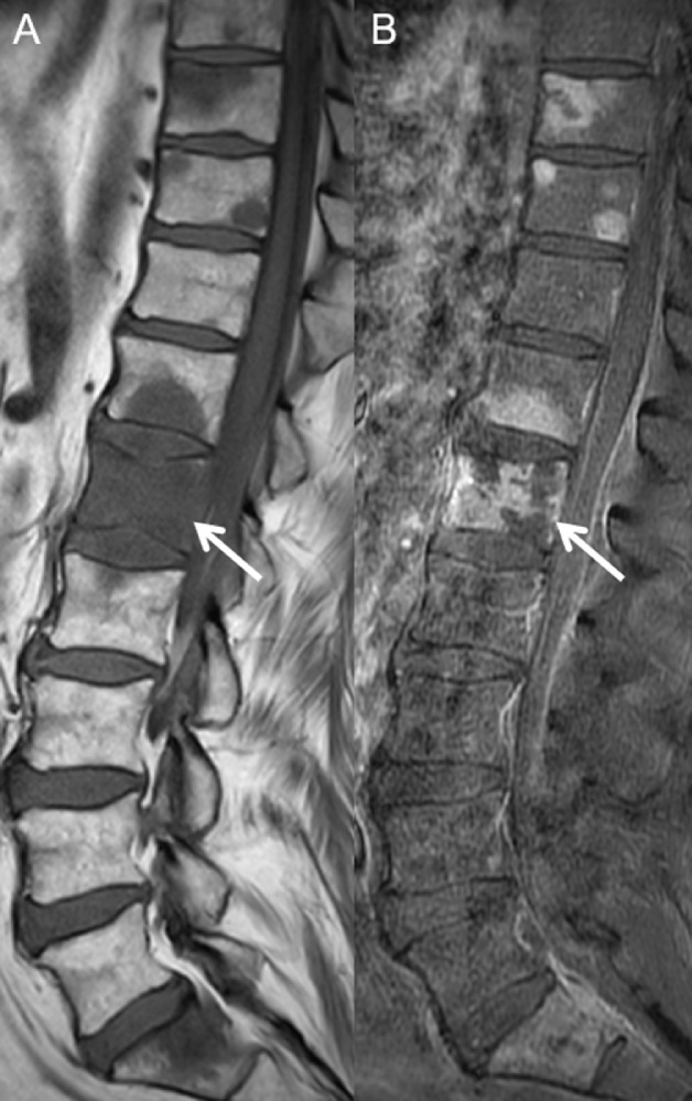
Spinal metastases are most commonly osseous and may extend to the epidural space. Less commonly, spinal metastases can be subdural, leptomeningeal, or intramedullary. Among these, subdural metastases are the most rare, with few reported cases. While these lesions are now almost exclusively detected on MRI, they can rarely be apparent on other modalities. It is important to recognize subdural metastases on any modality, because they have a significant impact on patient prognosis and treatment. We report a case of renal cell carcinoma in a 68-year-old male initially presenting with subdural metastases detected on CT myelography, with subsequent confirmation by MRI. The case illustrates, to our knowledge, the first example of subdural metastatic disease seen on CT myelography.
脊柱转移瘤最常见为骨转移,可延伸至硬膜外间隙。较少见的是,脊柱转移瘤可为硬膜下、软脑膜或髓内转移。其中,硬膜下转移最为罕见,报道病例极少。虽然现在这些病变几乎仅在磁共振成像(MRI)上被检测到,但在其他检查方式上很少能显影。在任何检查方式上识别硬膜下转移瘤都很重要,因为它们对患者的预后和治疗有重大影响。我们报告一例68岁男性肾细胞癌病例,最初在CT脊髓造影上发现硬膜下转移瘤,随后经MRI证实。据我们所知,该病例是CT脊髓造影上所见硬膜下转移瘤疾病的首个实例。