Wang Hongyun, Li Fengxia, Feng Jing, Wang Junping, Liu Xiaobing
Gastroenterology Department, Shanxi Provincial People's Hospital, Taiyuan, China.
Ann Transl Med. 2021 Apr;9(8):684. doi: 10.21037/atm-21-1070.
Peroxisome proliferators-activated receptors γ (PPARγ) and secreted frizzled related protein 5 (SFRP5) are abnormally expressed in liver cells. But their role in the transformation of non-alcoholic fatty liver (NAFL) to non-alcoholic steatohepatitis (NASH) remains to be studied. We aimed to explore the role of S-nitrosylation (SNO) in the conversion of NAFL to NASH via the peroxisome PPARγ/SFRP5 pathway.
A normal diet and methionine-choline deficient diet were used to construct the NAFL and NASH mouse models, respectively. The differences between the SNO of PPARγ in both models were measured by irreversible biotinylation. Quantitative reverse transcription PCR (qRT-PCR) and Western blotting were used to detect the effect of SNO on the expression of PPARγ messageRNA (mRNA) and protein in L02 hepatocytes. Nubiscan software, luciferase reporter gene, and chromatin immunoprecipitation assay (CHIP) were used to verify the targeting relationship between PPAR and SFRP5. The expression of tumor necrosis factor α (TNFα), interleukin-1β (IL-1β), and interleukin-6 (IL-6), which are indicators for the activation of Kupffer cells, were determined by enzyme linked immunosorbent assay (ELISA) after co-cultivation of L02 hepatocytes and Kupffer macrophages, as well as the exogenous regulation of SNO, PPARγ, and SFRP5 in hepatic L02 cells.
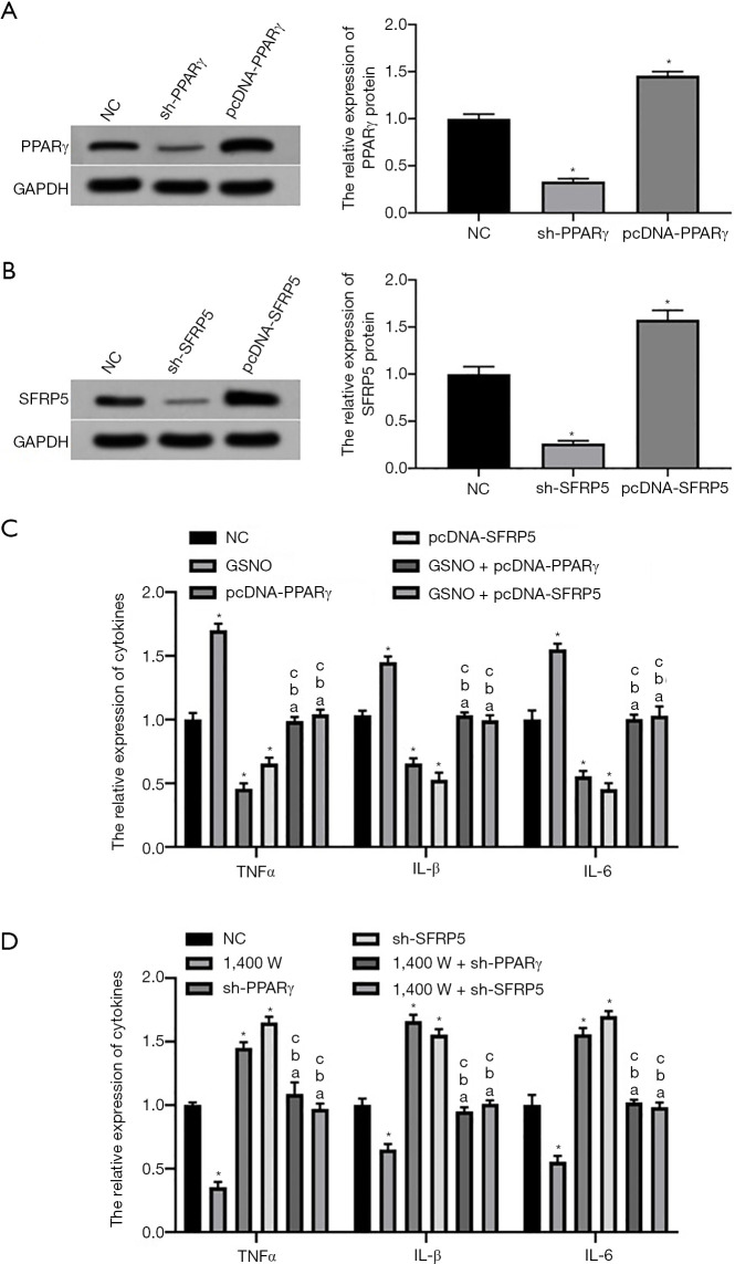
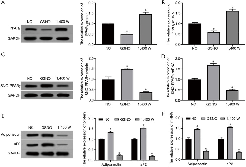
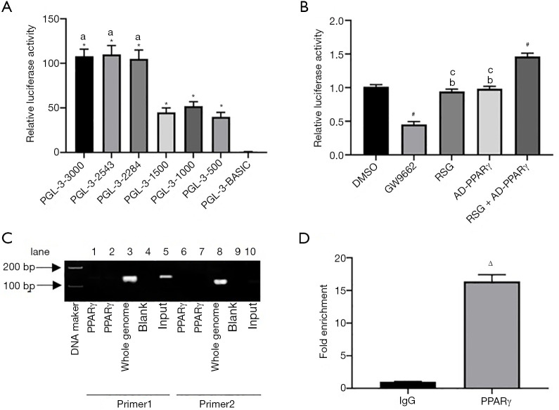
The NAFL and NASH mouse models were successfully constructed, and the level of PPARγ SNO in the NAFL model was significantly lower than the NASH model (P<0.05). The level of PPARγ was significantly downregulated after increasing the SNO of L02 cells, respectively (P<0.05). Nubiscan software and CHIP confirmed that PPARγ could bind to the promoter region of SFRP5 (P<0.05). Overexpression of PPARγ and SFRP5 could significantly downregulate the expression of TNFα, IL-1β, and IL-6 (P<0.05) correspondingly, while increasing the SNO level of L02 cells could restore the expression levels of TNFα, IL-1β, and IL-6.
SNO promoted the activation of macrophage Kupffer cells by inhibiting the PPARγ/SFRP5 pathway in L02 hepatocytes, thereby promoting the conversion of NAFL into NASH.
过氧化物酶体增殖物激活受体γ(PPARγ)和分泌型卷曲相关蛋白5(SFRP5)在肝细胞中异常表达。但其在非酒精性脂肪肝(NAFL)向非酒精性脂肪性肝炎(NASH)转变中的作用仍有待研究。我们旨在探讨S-亚硝基化(SNO)通过过氧化物酶体PPARγ/SFRP5途径在NAFL向NASH转化中的作用。
分别采用正常饮食和蛋氨酸-胆碱缺乏饮食构建NAFL和NASH小鼠模型。通过不可逆生物素化检测两种模型中PPARγ的SNO差异。采用定量逆转录PCR(qRT-PCR)和蛋白质印迹法检测SNO对L02肝细胞中PPARγ信使核糖核酸(mRNA)和蛋白质表达的影响。使用Nubiscan软件、荧光素酶报告基因和染色质免疫沉淀测定法(CHIP)验证PPAR与SFRP5之间的靶向关系。在L02肝细胞与库普弗巨噬细胞共培养以及对肝L02细胞中的SNO、PPARγ和SFRP5进行外源调节后,通过酶联免疫吸附测定法(ELISA)测定作为库普弗细胞激活指标的肿瘤坏死因子α(TNFα)、白细胞介素-1β(IL-1β)和白细胞介素-6(IL-6)的表达。
成功构建了NAFL和NASH小鼠模型,NAFL模型中PPARγ SNO水平显著低于NASH模型(P<0.05)。分别提高L02细胞的SNO后,PPARγ水平显著下调(P<0.05)。Nubiscan软件和CHIP证实PPARγ可与SFRP5启动子区域结合(P<0.05)。PPARγ和SFRP5的过表达可相应显著下调TNFα、IL-1β和IL-6的表达(P<0.05),而提高L02细胞的SNO水平可恢复TNFα、IL-1β和IL-6的表达水平。
SNO通过抑制L02肝细胞中的PPARγ/SFRP5途径促进巨噬细胞库普弗细胞的激活,从而促进NAFL向NASH的转化。