Laboratory of Optical and Molecular Imaging, Biomedical Engineering Institute, Polytechnique Montreal, Quebec, Canada.
Research Center, University Institute of Geriatrics of Montreal, Montreal, Quebec, Canada.
Hum Brain Mapp. 2021 Aug 15;42(12):3760-3776. doi: 10.1002/hbm.25463. Epub 2021 May 15.
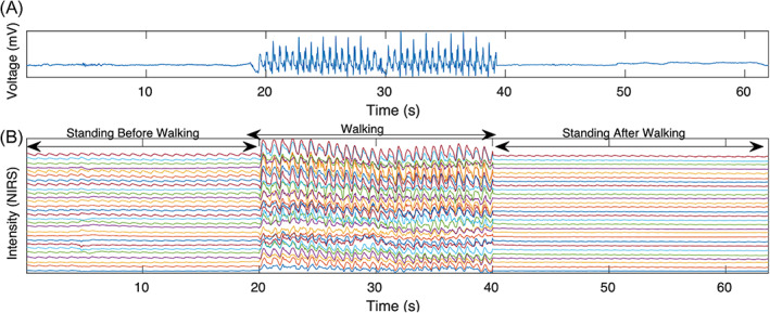
Recent studies have reported that optical indices of cerebral pulsatility are associated with cerebrovascular health in older adults. Such indices, including cerebral pulse amplitude and the pulse relaxation function (PRF), have been previously applied to quantify global and regional cerebral pulsatility. The aim of the present study was to determine whether these indices are modulated by cardiovascular status and whether they differ between individuals with low or high cardiovascular risk factors (LCVRF and HCVRF) and coronary artery disease (CAD). A total of 60 older adults aged 57-79 were enrolled in the study. Participants were grouped as LCVRF, HCVRF, and CAD. Participants were asked to walk freely on a gym track while a near-infrared spectroscopy (NIRS) device recorded hemodynamics data. Low-intensity, short-duration walking was used to test whether a brief cardiovascular challenge could increase the difference of pulsatility indices with respect to cardiovascular status. Results indicated that CAD individuals have higher global cerebral pulse amplitude compared with the other groups. Walking reduced global cerebral pulse amplitude and PRF in all groups but did not increase the difference across the groups. Instead, walking extended the spatial distribution of cerebral pulse amplitude to the anterior prefrontal cortex when CAD was compared to the CVRF groups. Further research is needed to determine whether cerebral pulse amplitude extracted from data acquired with NIRS, which is a noninvasive, inexpensive method, can provide an index to characterize the cerebrovascular status associated with CAD.
最近的研究报告称,大脑搏动的光学指数与老年人的脑血管健康有关。这些指数,包括大脑脉搏幅度和脉搏弛豫功能(PRF),之前曾被用于量化全局和局部大脑搏动。本研究的目的是确定这些指数是否受心血管状态的调节,以及它们在心血管危险因素低或高(LCVRF 和 HCVRF)和冠心病(CAD)个体之间是否存在差异。共有 60 名年龄在 57-79 岁的老年人参加了这项研究。参与者被分为 LCVRF、HCVRF 和 CAD 三组。要求参与者在健身房的轨道上自由行走,同时近红外光谱(NIRS)设备记录血液动力学数据。采用低强度、短时间的行走来测试短暂的心血管挑战是否会增加与心血管状态相关的搏动指数的差异。结果表明,与其他组相比,CAD 患者的全局大脑脉搏幅度更高。行走会降低所有组的全局大脑脉搏幅度和 PRF,但不会增加组间差异。相反,与 CVRF 组相比,当 CAD 与 CVRF 组相比时,行走会将大脑脉搏幅度的空间分布扩展到额前皮质。需要进一步研究,以确定从 NIRS 获得的数据中提取的大脑脉搏幅度是否可以作为一个非侵入性、成本低廉的方法来提供一个与 CAD 相关的脑血管状态的特征指数。