Diagnostic Radiology, Singapore General Hospital, Outram Road, Singapore, 169608, Singapore.
Duke-NUS Medical School, Singapore, Singapore.
Eur Radiol. 2021 Dec;31(12):9086-9097. doi: 10.1007/s00330-021-07871-4. Epub 2021 May 15.
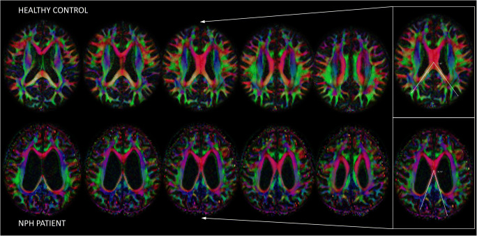
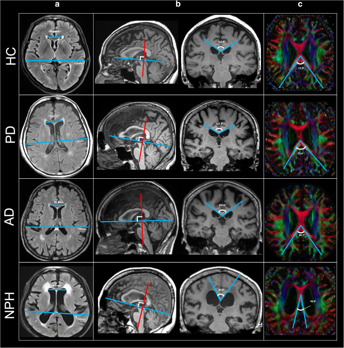
To evaluate the utility of the splenial angle (SA), an axial angular index of lateral ventriculomegaly measured on diffusion tensor MRI color fractional anisotropy maps, in differentiating NPH from Alzheimer's disease (AD), Parkinson's disease (PD), and healthy controls (HC), and post-shunt changes in NPH, compared to Evans' index and callosal angle.
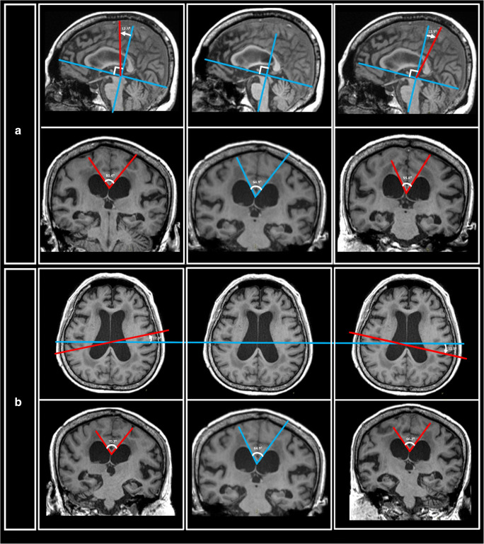
Evans' index, callosal angle, and SA were measured on brain MRI of 76 subjects comprising equal numbers of age- and sex-matched subjects from each cohort of NPH, AD, PD, and HC by two raters. Receiver operating characteristics (ROC) and multivariable analysis were used to assess the screening performance of each measure in differentiating and predicting NPH from non-NPH groups respectively. Temporal changes in the measures on 1-year follow-up MRI in 11 NPH patients (with or without ventriculoperitoneal shunting) were also assessed.
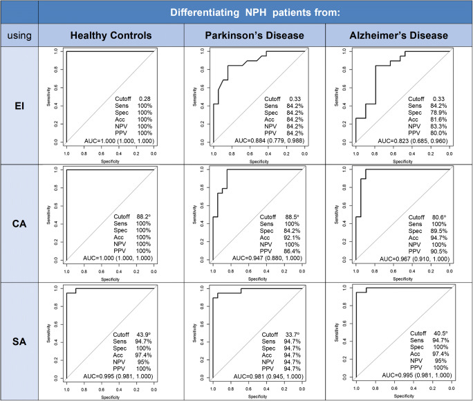
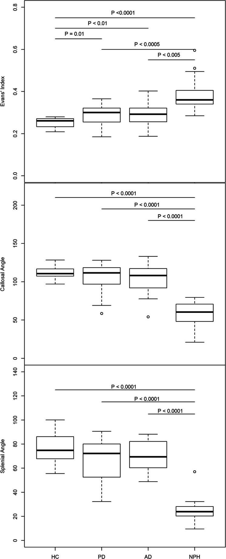
Inter-rater and intra-rater reliability were excellent for all measurements (intraclass correlation coefficients > 0.9). Pairwise comparison showed that SA was statistically different between NPH and AD/PD/HC subjects (p < 0.0001). SA performed the best in predicting NPH, with an area under the ROC curve of > 0.98, and was the only measure left in the final model of the multivariable analysis. Significant (p < 0.01) change in SA was seen at follow-up MRI of NPH patients who were shunted compared to those who were not.
The SA is readily measured on axial DTI color FA maps compared to the callosal angle and shows superior performance differentiating NPH from neurodegenerative disorders and sensitivity to ventricular changes in NPH after surgical intervention.
• The splenial angle is a novel simple angular radiological index proposed for idiopathic normal pressure hydrocephalus, measured in the ubiquitous axial plane on DTI color fractional anisotropy maps. • The splenial angle quantitates the compression and stretching of the posterior callosal commissural fibers alongside the distended lateral ventricles in idiopathic normal pressure hydrocephalus (NPH) using tools readily accessible in clinical practice and shows excellent test-retest reliability. • Splenial angle outperforms Evans' index and callosal angle in predicting NPH from healthy, Parkinson's disease, and Alzheimer's disease subjects on ROC analysis with an area under the curve of > 0.98 and is sensitive to morphological ventricular changes in NPH patients after ventricular shunting.
评估脑白质角(SA)在鉴别正常压力脑积水(NPH)与阿尔茨海默病(AD)、帕金森病(PD)和健康对照组(HC)中的效用,以及与 Evans 指数和胼胝体角相比,在 NPH 分流术后的变化。
由两位评分者在脑 MRI 上测量 76 名年龄和性别匹配的受试者的 Evans 指数、胼胝体角和 SA,这些受试者分别来自 NPH、AD、PD 和 HC 队列。使用受试者工作特征(ROC)曲线和多变量分析来评估每个指标在鉴别和预测 NPH 与非 NPH 组中的筛查性能。还评估了 11 例 NPH 患者(有或无脑室-腹腔分流术)在 1 年随访 MRI 上的测量值的时间变化。
所有测量的组内和组间可靠性均极佳(组内相关系数>0.9)。两两比较显示,SA 在 NPH 与 AD/PD/HC 受试者之间存在统计学差异(p<0.0001)。SA 在预测 NPH 方面表现最佳,ROC 曲线下面积>0.98,并且是多变量分析最终模型中唯一保留的指标。与未分流的 NPH 患者相比,接受分流术的 NPH 患者的 SA 在随访 MRI 上有显著变化(p<0.01)。
与胼胝体角相比,SA 更容易在轴向 DTI 彩色 FA 图谱上测量,并且在鉴别 NPH 与神经退行性疾病方面表现出更好的性能,并且对 NPH 手术后的脑室变化敏感。
脑白质角是一种用于特发性正常压力脑积水的新的简单角度放射学指标,在 DTI 彩色各向异性分数图谱的普遍轴向平面上进行测量。
脑白质角使用临床实践中易于获得的工具量化了特发性正常压力脑积水(NPH)中扩张的侧脑室旁的后连合纤维的压缩和拉伸,具有极好的测试-重测可靠性。
在 ROC 分析中,SA 在预测 NPH 与健康、帕金森病和阿尔茨海默病患者方面优于 Evans 指数和胼胝体角,曲线下面积>0.98,并且对 NPH 患者脑室分流术后的形态学脑室变化敏感。