• 文献检索
  • 文档翻译
  • 深度研究
  • 学术资讯
  • Suppr Zotero 插件Zotero 插件
  • 邀请有礼
  • 套餐&价格
  • 历史记录
应用&插件
Suppr Zotero 插件Zotero 插件浏览器插件Mac 客户端Windows 客户端微信小程序
定价
高级版会员购买积分包购买API积分包
服务
文献检索文档翻译深度研究API 文档MCP 服务
关于我们
关于 Suppr公司介绍联系我们用户协议隐私条款
关注我们

Suppr 超能文献

核心技术专利:CN118964589B侵权必究
粤ICP备2023148730 号-1Suppr @ 2026

文献检索

告别复杂PubMed语法,用中文像聊天一样搜索,搜遍4000万医学文献。AI智能推荐,让科研检索更轻松。

立即免费搜索

文件翻译

保留排版,准确专业,支持PDF/Word/PPT等文件格式,支持 12+语言互译。

免费翻译文档

深度研究

AI帮你快速写综述,25分钟生成高质量综述,智能提取关键信息,辅助科研写作。

立即免费体验

磁共振扩散加权成像在脑静脉血栓形成诊断中的应用:一项Meta分析

Diffusion-Weighted Magnetic Resonance Imaging in the Diagnosis of Cerebral Venous Thrombosis : A Meta-Analysis.

作者信息

Lv Bin, Jing Feng, Tian Cheng-Lin, Liu Jian-Chao, Wang Jun, Cao Xiang-Yu, Liu Xin-Feng, Yu Sheng-Yuan

机构信息

Department of Neurology, Chinese PLA General Hospital, Beijing, China.

Department of Medical Statistics, Chinese PLA General Hospital, Beijing, China.

出版信息

J Korean Neurosurg Soc. 2021 May;64(3):418-426. doi: 10.3340/jkns.2020.0247. Epub 2021 Apr 30.

DOI:10.3340/jkns.2020.0247
PMID:33993690
原文链接:https://pmc.ncbi.nlm.nih.gov/articles/PMC8128531/
Abstract

OBJECTIVE

A role of diffusion-weighted imaging (DWI) in the diagnosis of cerebral venous thrombosis (CVT) is not wellunderstood. This study evaluates the effectiveness of DWI in the diagnosis of CVT.

METHODS

Literature search was conducted in electronic databases for the identification of studies which reported the outcomes of patients subjected to DWI for CVT diagnosis. Random-effects meta-analyses were performed to achieve overall estimates of important diagnostic efficiency indices including hyperintense signal rate, the sensitivity and specificity of DWI in diagnosing CVT, and the apparent diffusion coefficient (ADC) of DWI signal areas and surrounding tissue.

RESULTS

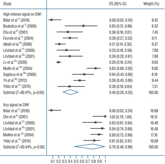
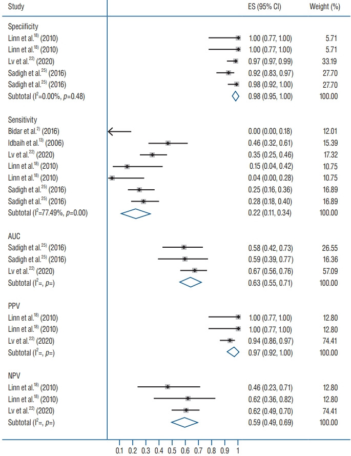
Nineteen studies (443 patients with 856 CVTs; age 40 years [95% confidence interval (CI), 33 to 43]; 28% males [95% CI, 18 to 38]; symptom onset to DWI time 4.6 days [95% CI, 2.3 to 6.9]) were included. Hyperintense signals on DWI were detected in 40% (95% CI, 26 to 55) of the cases. The sensitivity of DWI for detecting CVT was 22% (95% CI, 11 to 34) but specificity was 98% (95% CI, 95 to 100). ADC values were quite heterogenous in DWI signal areas. However, generally the ADC values were lower in DWI signal areas than in surrounding normal areas (mean difference-0.33×10⁻³ mm2/s [95% CI, -0.44 to -0.23]; p<0.00001).

CONCLUSION

DWI has a low sensitivity in detecting CVT and thus has a high risk of missing many CVT cases. However, because of its high specificity, it may have supporting and exploratory roles in CVT diagnosis.

摘要

目的

弥散加权成像(DWI)在脑静脉血栓形成(CVT)诊断中的作用尚未完全明确。本研究评估DWI在CVT诊断中的有效性。

方法

在电子数据库中进行文献检索,以识别报告了接受DWI进行CVT诊断的患者结果的研究。进行随机效应荟萃分析,以获得重要诊断效率指标的总体估计值,包括高信号率、DWI诊断CVT的敏感性和特异性,以及DWI信号区域和周围组织的表观扩散系数(ADC)。

结果

纳入19项研究(443例患者,共856处CVT;年龄40岁[95%置信区间(CI),33至43];28%为男性[95%CI,18至38];症状出现至进行DWI的时间为4.6天[95%CI,2.3至6.9])。40%(95%CI,26至55)的病例在DWI上检测到高信号。DWI检测CVT的敏感性为22%(95%CI,11至34),但特异性为98%(95%CI,95至100)。DWI信号区域的ADC值差异很大。然而,一般来说,DWI信号区域的ADC值低于周围正常区域(平均差异为-0.33×10⁻³mm2/s[95%CI,-0.44至-0.23];p<0.00001)。

结论

DWI在检测CVT方面敏感性较低,因此漏诊许多CVT病例的风险较高。然而,由于其高特异性,它可能在CVT诊断中具有辅助和探索作用。

https://cdn.ncbi.nlm.nih.gov/pmc/blobs/cda5/8128531/7cd60f77ac0f/jkns-2020-0247f5.jpg
https://cdn.ncbi.nlm.nih.gov/pmc/blobs/cda5/8128531/e0ba11aa303e/jkns-2020-0247f1.jpg
https://cdn.ncbi.nlm.nih.gov/pmc/blobs/cda5/8128531/a00b712e156f/jkns-2020-0247f2.jpg
https://cdn.ncbi.nlm.nih.gov/pmc/blobs/cda5/8128531/e29ac3ef70cf/jkns-2020-0247f3.jpg
https://cdn.ncbi.nlm.nih.gov/pmc/blobs/cda5/8128531/b8e688da99e2/jkns-2020-0247f4.jpg
https://cdn.ncbi.nlm.nih.gov/pmc/blobs/cda5/8128531/7cd60f77ac0f/jkns-2020-0247f5.jpg

相似文献

1
Diffusion-Weighted Magnetic Resonance Imaging in the Diagnosis of Cerebral Venous Thrombosis : A Meta-Analysis.磁共振扩散加权成像在脑静脉血栓形成诊断中的应用:一项Meta分析
J Korean Neurosurg Soc. 2021 May;64(3):418-426. doi: 10.3340/jkns.2020.0247. Epub 2021 Apr 30.
2
Role of diffusion-weighted imaging in the diagnosis of cerebral venous thrombosis.弥散加权成像在脑静脉血栓形成诊断中的作用
J Int Med Res. 2020 Jun;48(6):300060520933448. doi: 10.1177/0300060520933448.
3
Diffusion-weighted magnetic resonance in cerebral venous thrombosis.脑静脉血栓形成的磁共振扩散加权成像
Arch Neurol. 2001 Oct;58(10):1569-76. doi: 10.1001/archneur.58.10.1569.
4
Diffusion-Weighted MR Imaging Findings of Cortical Vein Thrombosis at 3 T.3T磁共振扩散加权成像对皮质静脉血栓形成的表现
Clin Neuroradiol. 2015 Sep;25(3):249-56. doi: 10.1007/s00062-014-0301-y. Epub 2014 Apr 5.
5
Diffusion- and perfusion-weighted magnetic resonance imaging in deep cerebral venous thrombosis.脑深部静脉血栓形成的扩散加权和灌注加权磁共振成像
Stroke. 1999 May;30(5):1144-6. doi: 10.1161/01.str.30.5.1144.
6
Diffusion-weighted imaging patterns of brain damage associated with cerebral venous thrombosis.与脑静脉血栓形成相关的脑损伤的弥散加权成像模式
AJNR Am J Neuroradiol. 2001 Feb;22(2):261-8.
7
[Diffusion-weighted magnetic resonance in deep cerebral venous thrombosis].[脑深部静脉血栓形成的磁共振扩散加权成像]
Neurologia. 2002 Oct;17(8):443-6.
8
Diffusion-weighted magnetic resonance imaging in superior sagittal sinus thrombosis.上矢状窦血栓形成的磁共振扩散加权成像
J Neuroimaging. 2002 Jul;12(3):267-9.
9
Perfusion and diffusion magnetic resonance imaging in human cerebral venous thrombosis.人脑静脉血栓形成的灌注与扩散磁共振成像
J Neurol. 2001 Jul;248(7):564-71. doi: 10.1007/s004150170133.
10
[Application of diffusion-weighted and perfusion magnetic resonance imaging in definition of the ischemic penumbra in hyperacute cerebral infarction].[弥散加权和灌注磁共振成像在超急性脑梗死缺血半暗带定义中的应用]
Zhonghua Yi Xue Za Zhi. 2003 Jun 10;83(11):952-7.

引用本文的文献

1
Imaging of Cerebral Venous Thrombosis.脑静脉血栓形成的影像学检查
Life (Basel). 2022 Aug 10;12(8):1215. doi: 10.3390/life12081215.

本文引用的文献

1
Role of diffusion-weighted imaging in the diagnosis of cerebral venous thrombosis.弥散加权成像在脑静脉血栓形成诊断中的作用
J Int Med Res. 2020 Jun;48(6):300060520933448. doi: 10.1177/0300060520933448.
2
Objective value on Apparent diffusion coefficient (ADC) map to categorize the intensity of diffusion-weighted imaging (DWI) restriction for prostate cancer detection on multiparametric prostate MRI.在多参数前列腺 MRI 上检测前列腺癌时,表观扩散系数 (ADC) 图上的目标值用于对扩散加权成像 (DWI) 受限的强度进行分类。
Int Braz J Urol. 2018 Sep-Oct;44(5):882-891. doi: 10.1590/S1677-5538.IBJU.2018.0038.
3
Clinical applications of diffusion weighted imaging in neuroradiology.
扩散加权成像在神经放射学中的临床应用。
Insights Imaging. 2018 Aug;9(4):535-547. doi: 10.1007/s13244-018-0624-3. Epub 2018 May 30.
4
Cerebral venous thrombosis.脑静脉血栓形成。
Nat Rev Neurol. 2017 Sep;13(9):555-565. doi: 10.1038/nrneurol.2017.104. Epub 2017 Aug 18.
5
Multiple Deep White Matter Hyperintense Lesions on Diffusion-Weighted Imaging: Early Sign of Straight Sinus Thrombosis.扩散加权成像上的多发深部白质高信号病变:直窦血栓形成的早期征象
J Stroke Cerebrovasc Dis. 2016 Sep;25(9):e131-3. doi: 10.1016/j.jstrokecerebrovasdis.2016.03.028. Epub 2016 Jul 18.
6
Assessment of cerebral venous sinus thrombosis using T2 (*)-weighted gradient echo magnetic resonance imaging sequences.使用T2(*)加权梯度回波磁共振成像序列评估脑静脉窦血栓形成。
Iran J Neurol. 2016 Apr 3;15(2):96-9.
7
Diagnostic Performance of MRI Sequences for Evaluation of Dural Venous Sinus Thrombosis.用于评估硬脑膜静脉窦血栓形成的MRI序列的诊断性能
AJR Am J Roentgenol. 2016 Jun;206(6):1298-306. doi: 10.2214/AJR.15.15719. Epub 2016 Mar 24.
8
Neuroimaging findings in pediatric cerebral sinovenous thrombosis.小儿脑静脉窦血栓形成的神经影像学表现。
Childs Nerv Syst. 2015 May;31(5):705-12. doi: 10.1007/s00381-015-2662-1. Epub 2015 Feb 26.
9
Diffusion-Weighted MR Imaging Findings of Cortical Vein Thrombosis at 3 T.3T磁共振扩散加权成像对皮质静脉血栓形成的表现
Clin Neuroradiol. 2015 Sep;25(3):249-56. doi: 10.1007/s00062-014-0301-y. Epub 2014 Apr 5.
10
Diagnosis and treatment of cerebral venous thrombosis.脑静脉血栓形成的诊断与治疗
Expert Rev Cardiovasc Ther. 2012 Dec;10(12):1545-53. doi: 10.1586/erc.12.126.