Drake-Pérez Marta, Boto Jose, Fitsiori Aikaterini, Lovblad Karl, Vargas Maria Isabel
Division of Diagnostic and Interventional Neuroradiology of Geneva University Hospitals, DISIM and Faculty of Medicine of Geneva, Rue Gabrielle-Perret-Gentil 4, 1211, Genève 14, Switzerland.
Department of Radiology, University Hospital Marqués de Valdecilla - IDIVAL, Santander, Spain.
Insights Imaging. 2018 Aug;9(4):535-547. doi: 10.1007/s13244-018-0624-3. Epub 2018 May 30.
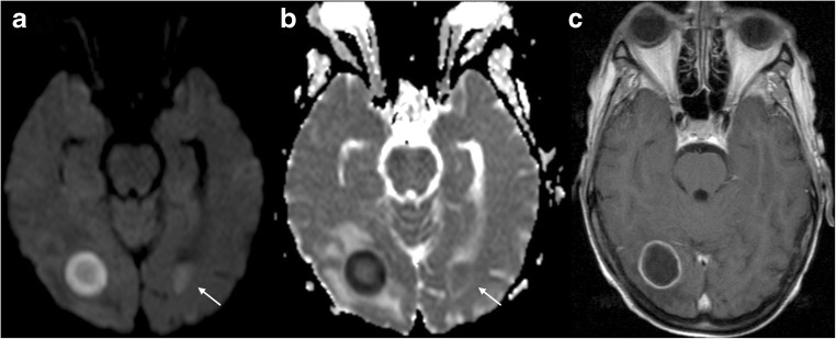
Diffusion-weighted imaging (DWI) has revolutionised stroke imaging since its introduction in the mid-1980s, and it has also become a pillar of current neuroimaging. Diffusion abnormalities represent alterations in the random movement of water molecules in tissues, revealing their microarchitecture, and occur in many neurological conditions. DWI provides useful information, increasing the sensitivity of MRI as a diagnostic tool, narrowing the differential diagnosis, providing prognostic information, aiding in treatment planning and evaluating response to treatment. Recently, there have been several technical improvements in DWI, leading to reduced acquisition time and artefacts and enabling the development of diffusion tensor imaging (DTI) as a tool for assessing white matter. We aim to review the main clinical uses of DWI, focusing on the physiological mechanisms that lead to diffusion abnormalities. Common pitfalls will also be addressed.
• DWI includes EPI, TSE, RESOLVE or EPI combined with reduced volume excitation. • DWI is the most sensitive sequence in stroke diagnosis and provides information about prognosis. • DWI helps in the detection of intramural haematomas (arterial dissection). • In diffusion imaging, ADC is inversely proportional to tumour cellularity. • DWI and DTI derived parameters can be used as biomarkers in different pathologies.
自20世纪80年代中期引入以来,扩散加权成像(DWI)彻底改变了中风成像,并且它也已成为当前神经成像的支柱。扩散异常代表组织中水分子随机运动的改变,揭示其微观结构,并且发生在许多神经系统疾病中。DWI提供有用信息,提高了MRI作为诊断工具的敏感性,缩小了鉴别诊断范围,提供了预后信息,有助于治疗计划制定并评估治疗反应。最近,DWI有了几项技术改进,导致采集时间和伪影减少,并使扩散张量成像(DTI)得以发展成为评估白质的工具。我们旨在回顾DWI的主要临床应用,重点关注导致扩散异常的生理机制。还将讨论常见的陷阱。
• DWI包括回波平面成像(EPI)、快速自旋回波(TSE)、分辨率优化视图采集(RESOLVE)或EPI结合减少容积激发。• DWI是中风诊断中最敏感序列,并提供预后信息。• DWI有助于检测壁内血肿(动脉夹层)。• 在扩散成像中,表观扩散系数(ADC)与肿瘤细胞密度成反比。• DWI和DTI衍生参数可在不同病理中用作生物标志物。