De-Miguel Francisco F, Leon-Pinzon Carolina, Torres-Platas Susana G, Del-Pozo Vanessa, Hernández-Mendoza Guillermo A, Aguirre-Olivas Dilia, Méndez Bruno, Moore Sharlen, Sánchez-Sugía Celeste, García-Aguilera Marco Antonio, Martínez-Valencia Alejandro, Ramírez-Santiago Guillermo, Rubí J Miguel
Instituto de Fisiología Celular-Neurociencias, Universidad Nacional Autónoma de México, México City, Mexico.
Centro de Ciencias de la Complejidad, Universidad Nacional Autónoma de México, México City, Mexico.
Front Mol Neurosci. 2021 Apr 30;14:638858. doi: 10.3389/fnmol.2021.638858. eCollection 2021.
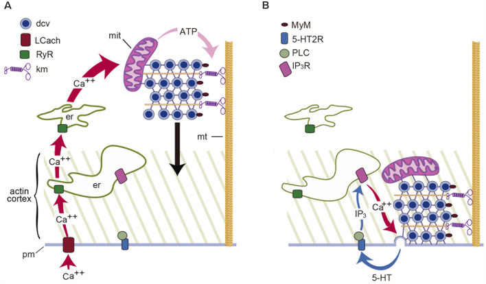
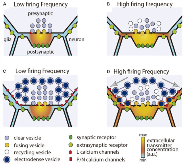
Streams of action potentials or long depolarizations evoke a massive exocytosis of transmitters and peptides from the surface of dendrites, axons and cell bodies of different neuron types. Such mode of exocytosis is known as extrasynaptic for occurring without utilization of synaptic structures. Most transmitters and all peptides can be released extrasynaptically. Neurons may discharge their contents with relative independence from the axon, soma and dendrites. Extrasynaptic exocytosis takes fractions of a second in varicosities or minutes in the soma or dendrites, but its effects last from seconds to hours. Unlike synaptic exocytosis, which is well localized, extrasynaptic exocytosis is diffuse and affects neuronal circuits, glia and blood vessels. Molecules that are liberated may reach extrasynaptic receptors microns away. The coupling between excitation and exocytosis follows a multistep mechanism, different from that at synapses, but similar to that for the release of hormones. The steps from excitation to exocytosis have been studied step by step for the vital transmitter serotonin in leech Retzius neurons. The events leading to serotonin exocytosis occur similarly for the release of other transmitters and peptides in central and peripheral neurons. Extrasynaptic exocytosis occurs commonly onto glial cells, which react by releasing the same or other transmitters. In the last section, we discuss how illumination of the retina evokes extrasynaptic release of dopamine and ATP. Dopamine contributes to light-adaptation; ATP activates glia, which mediates an increase in blood flow and oxygenation. A proper understanding of the workings of the nervous system requires the understanding of extrasynaptic communication.
一连串的动作电位或长时间的去极化会引发不同类型神经元的树突、轴突和细胞体表面大量释放递质和肽。这种胞吐作用模式因不利用突触结构而发生,被称为非突触性胞吐。大多数递质和所有肽都可以通过非突触方式释放。神经元可以相对独立地从轴突、胞体和树突释放其内容物。非突触性胞吐在曲张体中只需几分之一秒,在胞体或树突中则需要几分钟,但其影响可持续数秒至数小时。与定位良好的突触性胞吐不同,非突触性胞吐是弥散性的,会影响神经回路、神经胶质细胞和血管。释放的分子可能会到达数微米外的非突触受体。兴奋与胞吐之间的偶联遵循一种多步骤机制,不同于突触处的机制,但类似于激素释放的机制。对于水蛭Retzius神经元中的重要递质5-羟色胺,已经逐步研究了从兴奋到胞吐的各个步骤。导致5-羟色胺胞吐的事件在中枢和外周神经元中其他递质和肽的释放过程中也类似发生。非突触性胞吐通常发生在神经胶质细胞上,神经胶质细胞会通过释放相同或其他递质做出反应。在最后一部分,我们讨论视网膜光照如何引发多巴胺和ATP的非突触性释放。多巴胺有助于光适应;ATP激活神经胶质细胞,后者介导血流量和氧合作用的增加。对神经系统工作方式的正确理解需要了解非突触性通讯。