Trueta Citlali, De-Miguel Francisco F
Departamento de Neurofisiología, Instituto Nacional de Psiquiatría Ramón de la Fuente Muñiz México, D.F., México.
Front Physiol. 2012 Sep 4;3:319. doi: 10.3389/fphys.2012.00319. eCollection 2012.
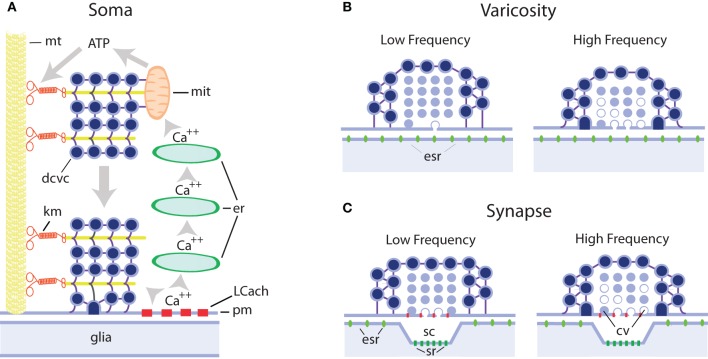
We review the evidence of exocytosis from extrasynaptic sites in the soma, dendrites, and axonal varicosities of central and peripheral neurons of vertebrates and invertebrates, with emphasis on somatic exocytosis, and how it contributes to signaling in the nervous system. The finding of secretory vesicles in extrasynaptic sites of neurons, the presence of signaling molecules (namely transmitters or peptides) in the extracellular space outside synaptic clefts, and the mismatch between exocytosis sites and the location of receptors for these molecules in neurons and glial cells, have long suggested that in addition to synaptic communication, transmitters are released, and act extrasynaptically. The catalog of these molecules includes low molecular weight transmitters such as monoamines, acetylcholine, glutamate, gama-aminobutiric acid (GABA), adenosine-5-triphosphate (ATP), and a list of peptides including substance P, brain-derived neurotrophic factor (BDNF), and oxytocin. By comparing the mechanisms of extrasynaptic exocytosis of different signaling molecules by various neuron types we show that it is a widespread mechanism for communication in the nervous system that uses certain common mechanisms, which are different from those of synaptic exocytosis but similar to those of exocytosis from excitable endocrine cells. Somatic exocytosis has been measured directly in different neuron types. It starts after high-frequency electrical activity or long experimental depolarizations and may continue for several minutes after the end of stimulation. Activation of L-type calcium channels, calcium release from intracellular stores and vesicle transport towards the plasma membrane couple excitation and exocytosis from small clear or large dense core vesicles in release sites lacking postsynaptic counterparts. The presence of synaptic and extrasynaptic exocytosis endows individual neurons with a wide variety of time- and space-dependent communication possibilities. Extrasynaptic exocytosis may be the major source of signaling molecules producing volume transmission and by doing so may be part of a long duration signaling mode in the nervous system.
我们回顾了脊椎动物和无脊椎动物中枢及外周神经元的胞体、树突和轴突曲张体中突触外位点胞吐作用的证据,重点关注体细胞胞吐作用,以及它如何促进神经系统中的信号传导。在神经元的突触外位点发现分泌囊泡,在突触间隙外的细胞外空间存在信号分子(即递质或肽),以及胞吐作用位点与这些分子在神经元和神经胶质细胞中的受体位置不匹配,长期以来表明除了突触通讯外,递质也会释放并在突触外起作用。这些分子的目录包括低分子量递质,如单胺、乙酰胆碱、谷氨酸、γ-氨基丁酸(GABA)、三磷酸腺苷(ATP),以及一系列肽,包括P物质、脑源性神经营养因子(BDNF)和催产素。通过比较不同神经元类型对不同信号分子的突触外胞吐作用机制,我们表明这是神经系统中一种广泛的通讯机制,它使用某些共同机制,这些机制不同于突触胞吐作用,但类似于可兴奋内分泌细胞的胞吐作用。已经在不同类型的神经元中直接测量了体细胞胞吐作用。它在高频电活动或长时间实验性去极化后开始,并且在刺激结束后可能持续几分钟。L型钙通道的激活、细胞内钙库的钙释放以及囊泡向质膜的转运,将兴奋与缺乏突触后对应物的释放位点中小而清亮或大而致密核心囊泡的胞吐作用耦合起来。突触和突触外胞吐作用的存在赋予单个神经元广泛的时间和空间依赖性通讯可能性。突触外胞吐作用可能是产生容积传递的信号分子的主要来源,通过这样做可能是神经系统中长时程信号模式的一部分。