Sun Ke, Yu Tao, Yang Dongju, Ren Zhiwei, Qiao Liang, Ni Duanyu, Wang Xueyuan, Zhao Yongxiang, Chen Xin, Xiang Jing, Chen Nan, Gao Runshi, Yang Kun, Lin Yicong, Kober Tobias, Zhang Guojun
Beijing Institute of Functional Neurosurgery, Xuanwu Hospital, Capital Medical University, Beijing, China.
Department of Neurology, Xuanwu Hospital, Capital Medical University, Beijing, China.
Front Neurol. 2021 Apr 29;12:651592. doi: 10.3389/fneur.2021.651592. eCollection 2021.
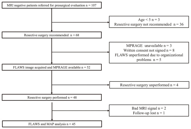
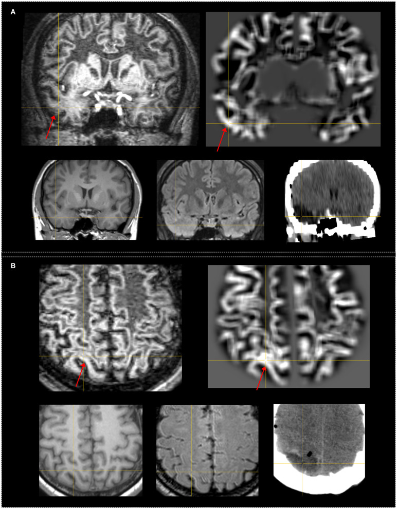
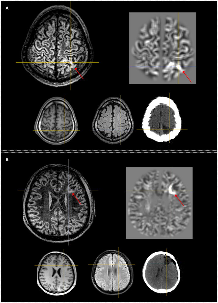
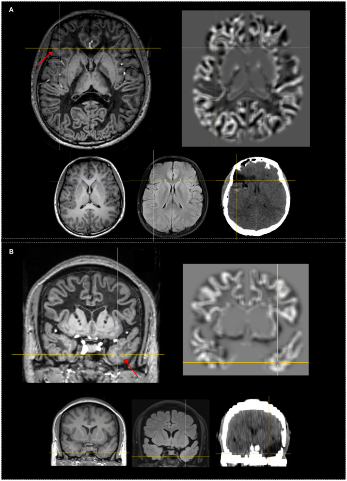
Delineation of subtle lesions in magnetic resonance imaging (MRI)-negative patients is of great importance in preoperative epilepsy evaluation. The aim of our study was to explore the diagnostic value of the novel fluid and white matter suppression (FLAWS) sequence in comparison with a voxel-based MRI postprocessing morphometric analysis program (MAP) in a consecutive cohort of non-lesional patients. Surgical candidates with a negative finding on an official neuroradiology report were enrolled. High-resolution FLAWS image and MAP maps generated based on high-resolution three-dimensional (3D) T1 image were visually inspected for each patient. The findings of FLAWS or MAP-positive (FLAWS/MAP+) regions were compared with the surgical resection cavity in correlation with surgical outcome and pathology. Forty-five patients were enrolled; the pathological examination revealed focal cortical dysplasia (FCD) in 32 patients and other findings in 13 patients. The positive rate, sensitivity, and specificity were 48.9%, 0.43, and 0.87, respectively, for FLAWS and 64.4%, 0.57, and 0.8, respectively, for MAP. Concordance between surgical resection and FLAWS+ or MAP+ regions was significantly associated with a seizure-free outcome (FLAWS: = 0.002; MAP: = 0.0003). A positive finding in FLAWS and MAP together with abnormalities in the same gyrus (FLAWS-MAP gyral+) was detected in 31.1% of patients. FLAWS+ only and MAP+ only were found in 7 (15.5%) and 14 (31.1%) patients, respectively. FLAWS showed a promising value for identifying subtle epileptogenic lesions and can be used as a complement to current MAP in patients with MRI-negative epilepsy.
在术前癫痫评估中,明确磁共振成像(MRI)检查结果为阴性的患者体内的细微病变至关重要。本研究旨在探讨新型液体与白质抑制(FLAWS)序列与基于体素的MRI后处理形态计量分析程序(MAP)相比,在连续队列的非病变患者中的诊断价值。纳入官方神经放射学报告结果为阴性的手术候选患者。对每位患者的高分辨率FLAWS图像以及基于高分辨率三维(3D)T1图像生成的MAP图谱进行视觉检查。将FLAWS或MAP阳性(FLAWS/MAP+)区域的检查结果与手术切除腔进行比较,并与手术结果和病理情况相关联。共纳入45例患者;病理检查显示32例患者存在局灶性皮质发育不良(FCD),13例患者有其他检查结果。FLAWS的阳性率、敏感性和特异性分别为48.9%、0.43和0.87,MAP的阳性率、敏感性和特异性分别为64.4%、0.57和0.8。手术切除与FLAWS+或MAP+区域之间的一致性与无癫痫发作的结果显著相关(FLAWS:=0.002;MAP:=0.0003)。31.1%的患者在FLAWS和MAP检查中均呈阳性,且同一脑回存在异常(FLAWS-MAP脑回+)。仅FLAWS+和仅MAP+分别在7例(15.5%)和14例(31.1%)患者中发现。FLAWS在识别细微癫痫病灶方面显示出有前景的价值,可作为当前MRI阴性癫痫患者MAP检查的补充手段。