• 文献检索
  • 文档翻译
  • 深度研究
  • 学术资讯
  • Suppr Zotero 插件Zotero 插件
  • 邀请有礼
  • 套餐&价格
  • 历史记录
应用&插件
Suppr Zotero 插件Zotero 插件浏览器插件Mac 客户端Windows 客户端微信小程序
定价
高级版会员购买积分包购买API积分包
服务
文献检索文档翻译深度研究API 文档MCP 服务
关于我们
关于 Suppr公司介绍联系我们用户协议隐私条款
关注我们

Suppr 超能文献

核心技术专利:CN118964589B侵权必究
粤ICP备2023148730 号-1Suppr @ 2026

文献检索

告别复杂PubMed语法,用中文像聊天一样搜索,搜遍4000万医学文献。AI智能推荐,让科研检索更轻松。

立即免费搜索

文件翻译

保留排版,准确专业,支持PDF/Word/PPT等文件格式,支持 12+语言互译。

免费翻译文档

深度研究

AI帮你快速写综述,25分钟生成高质量综述,智能提取关键信息,辅助科研写作。

立即免费体验

暴露磷脂酰丝氨酸的肿瘤源性微粒会加剧三阴性乳腺癌中的凝血和癌细胞跨内皮迁移。

Phosphatidylserine-exposing tumor-derived microparticles exacerbate coagulation and cancer cell transendothelial migration in triple-negative breast cancer.

作者信息

Zhang Cong, Yang Zhuowen, Zhou Peng, Yu Muxin, Li Baorong, Liu Yingmiao, Jin Jiaqi, Liu Wenhui, Jing Haijiao, Du Jingwen, Tian Jie, Zhao Zhiyu, Wang Jianxin, Chu Yinzhu, Zhang ChunMei, Novakovic Valerie A, Shi Jialan, Wu Changjun

机构信息

Department of Ultrasound, The First Hospital, Harbin Medical University, Harbin, China.

Department of Gerontology, The First Hospital, Harbin Medical University, Harbin, China.

出版信息

Theranostics. 2021 Apr 19;11(13):6445-6460. doi: 10.7150/thno.53637. eCollection 2021.

DOI:10.7150/thno.53637
PMID:33995667
原文链接:https://pmc.ncbi.nlm.nih.gov/articles/PMC8120203/
Abstract

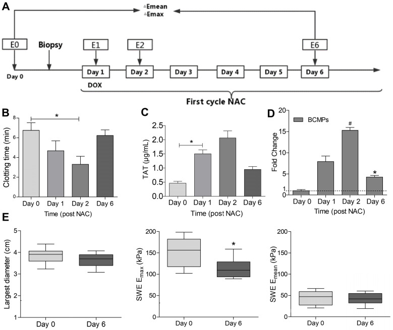
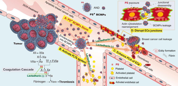
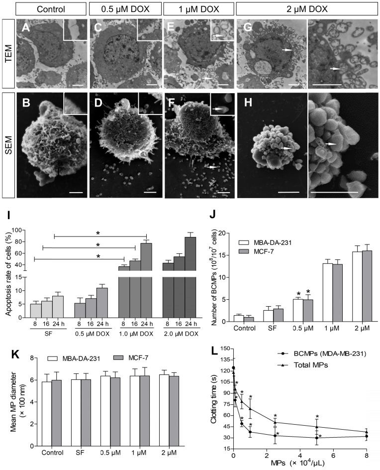
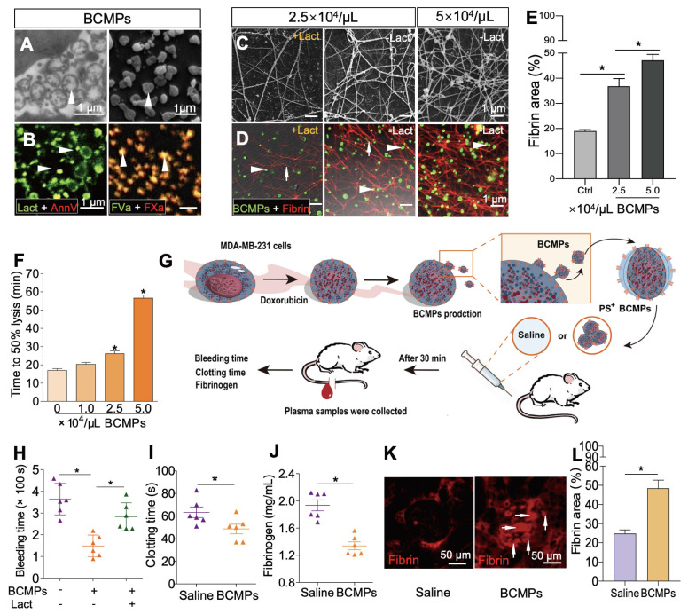
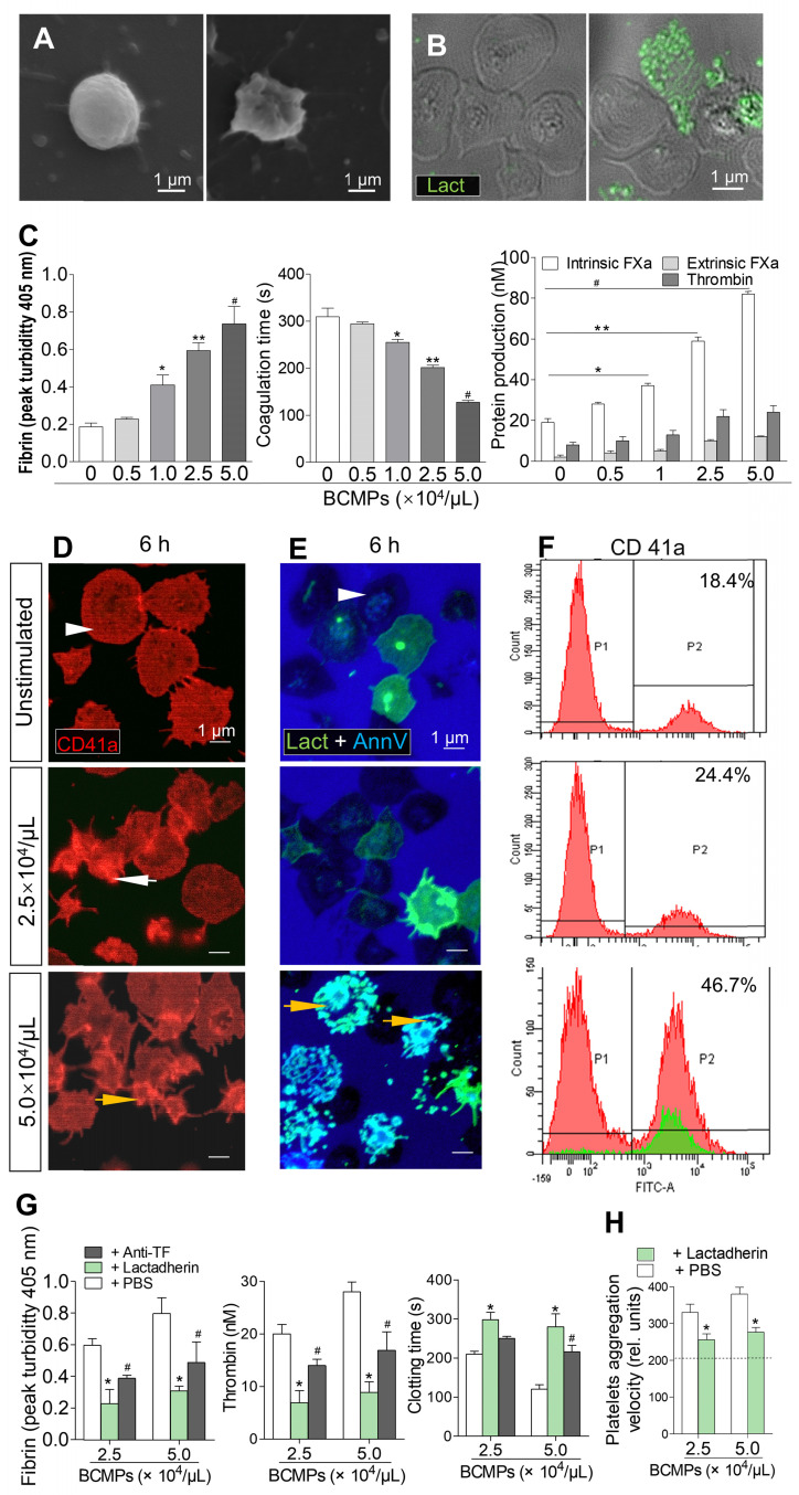
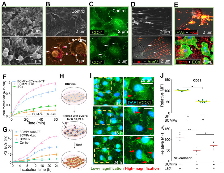
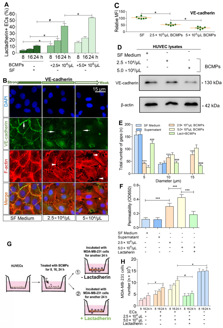
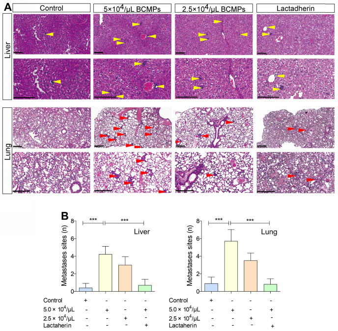
Neoadjuvant chemotherapy is relevant to the formation of thromboembolism and secondary neoplasms in triple-negative breast cancer (TNBC). Chemotherapy-induced breast cancer cell-derived microparticles (BCMPs) may have important thrombogenic and pro-metastatic effects on platelets and endothelium, which may be related to the expression and distribution of phosphatidylserine (PS). However, investigating these interactions is challenging due to technical limitations. A study was conducted in 20 healthy individuals and 18 patients who had been recently diagnosed with TNBC and were undergoing neoadjuvant chemotherapy with doxorubicin and cyclophosphamide. BCMPs were isolated from patient blood samples and doxorubicin-treated breast cancer cell lines. Their structure and morphology were studied by electron microscopy and antigen levels were measured by fluorescence-activated cell sorting. In an inhibition assay, isolated BCMPs were pretreated with lactadherin or tissue factor antibodies. Platelets isolated from healthy subjects were treated with BCMPs and coagulation time, fibrin formation, and expression of intrinsic/extrinsic factor Xase (FXa) and thrombin were evaluated. The effects of BCMPs on endothelial thrombogenicity and integrity were assessed by confocal microscopy, electron microscopy, measurement of intrinsic/extrinsic FXa, prothrombinase assay, and transwell permeability assay. Neoadjuvant chemotherapy significantly increased the expression of PS+ BCMPs in patient plasma. Its expression was associated with a rapid increase in procoagulant activity. Treatment with lactadherin, a PS-binding scavenging molecule, markedly reduced the adhesion of BCMPs and abolished their procoagulant activity, but this was not observed with tissue factor antibody treatment. Intravenous injection of BCMPs in mice induced a significant hypercoagulable state, reducing the extent of plasma fibrinogen and promoting the appearance of new thrombus. Cancer cells incubated with doxorubicin released large numbers of PS+ BCMPs, which stimulated and transformed endothelial cells into a procoagulant phenotype and increased the aggregation and activation of platelets. Moreover, cancer cells exploited this BCMP-induced endothelial leakiness and showed promoted metastasis. Pretreatment with lactadherin increased uptake of both PS+ BCMPs and cancer cells by endothelial cells and limited the transendothelial migration of cancer cells. Lactadherin, a biosensor that we developed, was used to study the extracellular vesicle distribution of PS, which revealed a novel PS+ BCMPs administrative axis that initiated a local coagulation cascade and facilitated metastatic colonization of circulating cancer cells.

摘要

新辅助化疗与三阴性乳腺癌(TNBC)中血栓栓塞和继发性肿瘤的形成有关。化疗诱导的乳腺癌细胞衍生微粒(BCMPs)可能对血小板和内皮细胞具有重要的促血栓形成和促转移作用,这可能与磷脂酰丝氨酸(PS)的表达和分布有关。然而,由于技术限制,研究这些相互作用具有挑战性。对20名健康个体和18名最近被诊断为TNBC并正在接受阿霉素和环磷酰胺新辅助化疗的患者进行了一项研究。从患者血液样本和阿霉素处理的乳腺癌细胞系中分离出BCMPs。通过电子显微镜研究其结构和形态,并通过荧光激活细胞分选测量抗原水平。在抑制试验中,用乳黏附素或组织因子抗体预处理分离出的BCMPs。用BCMPs处理从健康受试者分离出的血小板,并评估凝血时间、纤维蛋白形成以及内源性/外源性因子Xa(FXa)和凝血酶的表达。通过共聚焦显微镜、电子显微镜、内源性/外源性FXa的测量、凝血酶原酶测定和Transwell通透性测定评估BCMPs对内皮血栓形成性和完整性的影响。新辅助化疗显著增加了患者血浆中PS+ BCMPs的表达。其表达与促凝活性的快速增加有关。用PS结合清除分子乳黏附素治疗可显著降低BCMPs的黏附并消除其促凝活性,但组织因子抗体治疗未观察到这种情况。给小鼠静脉注射BCMPs会诱导显著的高凝状态,降低血浆纤维蛋白原水平并促进新血栓的出现。用阿霉素孵育的癌细胞释放大量PS+ BCMPs,其刺激内皮细胞并将其转化为促凝表型,增加血小板的聚集和活化。此外,癌细胞利用这种BCMP诱导的内皮渗漏并显示出转移促进作用。用乳黏附素预处理可增加内皮细胞对PS+ BCMPs和癌细胞的摄取,并限制癌细胞的跨内皮迁移。我们开发的生物传感器乳黏附素用于研究PS的细胞外囊泡分布,揭示了一个新的PS+ BCMPs管理轴,该轴启动局部凝血级联反应并促进循环癌细胞的转移定植。

https://cdn.ncbi.nlm.nih.gov/pmc/blobs/b2fc/8120203/f42270d86c42/thnov11p6445g008.jpg
https://cdn.ncbi.nlm.nih.gov/pmc/blobs/b2fc/8120203/5dd71ff732d9/thnov11p6445g001.jpg
https://cdn.ncbi.nlm.nih.gov/pmc/blobs/b2fc/8120203/bfb14aba16dc/thnov11p6445g002.jpg
https://cdn.ncbi.nlm.nih.gov/pmc/blobs/b2fc/8120203/ce7d767a2942/thnov11p6445g003.jpg
https://cdn.ncbi.nlm.nih.gov/pmc/blobs/b2fc/8120203/682059b8c31b/thnov11p6445g004.jpg
https://cdn.ncbi.nlm.nih.gov/pmc/blobs/b2fc/8120203/9183854d5475/thnov11p6445g005.jpg
https://cdn.ncbi.nlm.nih.gov/pmc/blobs/b2fc/8120203/fc9bfd44792d/thnov11p6445g006.jpg
https://cdn.ncbi.nlm.nih.gov/pmc/blobs/b2fc/8120203/da8eed3d364a/thnov11p6445g007.jpg
https://cdn.ncbi.nlm.nih.gov/pmc/blobs/b2fc/8120203/f42270d86c42/thnov11p6445g008.jpg

相似文献

1
Phosphatidylserine-exposing tumor-derived microparticles exacerbate coagulation and cancer cell transendothelial migration in triple-negative breast cancer.暴露磷脂酰丝氨酸的肿瘤源性微粒会加剧三阴性乳腺癌中的凝血和癌细胞跨内皮迁移。
Theranostics. 2021 Apr 19;11(13):6445-6460. doi: 10.7150/thno.53637. eCollection 2021.
2
Phosphatidylserine exposing-platelets and microparticles promote procoagulant activity in colon cancer patients.暴露磷脂酰丝氨酸的血小板和微粒促进结肠癌患者的促凝活性。
J Exp Clin Cancer Res. 2016 Mar 25;35:54. doi: 10.1186/s13046-016-0328-9.
3
Increased blood cell phosphatidylserine exposure and circulating microparticles contribute to procoagulant activity after carotid artery stenting.血小板磷脂酰丝氨酸暴露增加和循环微颗粒导致颈动脉支架置入术后促凝活性增强。
J Neurosurg. 2017 Nov;127(5):1041-1054. doi: 10.3171/2016.8.JNS16996. Epub 2016 Dec 23.
4
Phosphotidylserine exposure and neutrophil extracellular traps enhance procoagulant activity in patients with inflammatory bowel disease.磷脂酰丝氨酸暴露和中性粒细胞胞外陷阱增强炎症性肠病患者的促凝血活性。
Thromb Haemost. 2016 Apr;115(4):738-51. doi: 10.1160/TH15-09-0710. Epub 2015 Dec 10.
5
Intravascular cells and circulating microparticles induce procoagulant activity via phosphatidylserine exposure in heart failure.血管内细胞和循环微颗粒通过心力衰竭中的磷脂酰丝氨酸暴露诱导促凝活性。
J Thromb Thrombolysis. 2019 Aug;48(2):187-194. doi: 10.1007/s11239-019-01889-8.
6
CIRCULATING MICROPARTICLES, BLOOD CELLS, AND ENDOTHELIUM INDUCE PROCOAGULANT ACTIVITY IN SEPSIS THROUGH PHOSPHATIDYLSERINE EXPOSURE.循环微粒、血细胞和内皮细胞通过磷脂酰丝氨酸暴露在脓毒症中诱导促凝活性。
Shock. 2016 Mar;45(3):299-307. doi: 10.1097/SHK.0000000000000509.
7
Increased phosphatidylserine-exposing microparticles and their originating cells are associated with the coagulation process in patients with IgA nephropathy.在 IgA 肾病患者中,暴露磷脂酰丝氨酸的微粒及其来源细胞与凝血过程有关。
Nephrol Dial Transplant. 2016 May;31(5):747-59. doi: 10.1093/ndt/gfv403. Epub 2015 Dec 15.
8
Phosphatidylserine on microparticles and associated cells contributes to the hypercoagulable state in diabetic kidney disease.微粒体和相关细胞上的磷脂酰丝氨酸导致糖尿病肾病的高凝状态。
Nephrol Dial Transplant. 2018 Dec 1;33(12):2115-2127. doi: 10.1093/ndt/gfy027.
9
Phosphatidylserine on blood cells and endothelial cells contributes to the hypercoagulable state in cirrhosis.血细胞和内皮细胞上的磷脂酰丝氨酸导致肝硬化中的高凝状态。
Liver Int. 2016 Dec;36(12):1800-1810. doi: 10.1111/liv.13167. Epub 2016 Jun 22.
10
Microparticles and blood cells induce procoagulant activity via phosphatidylserine exposure in NSTEMI patients following stent implantation.在支架植入后的非ST段抬高型心肌梗死患者中,微粒和血细胞通过磷脂酰丝氨酸暴露诱导促凝活性。
Int J Cardiol. 2016 Nov 15;223:121-128. doi: 10.1016/j.ijcard.2016.07.260. Epub 2016 Aug 2.

引用本文的文献

1
Apoptotic cells promote circulating tumor cell survival and metastasis.凋亡细胞促进循环肿瘤细胞的存活和转移。
Commun Biol. 2025 Jul 29;8(1):1121. doi: 10.1038/s42003-025-08541-7.
2
Elucidating stroke etiology through lipidomics of thrombi and plasma in acute ischemic stroke patients undergoing endovascular thrombectomy.通过对接受血管内血栓切除术的急性缺血性中风患者的血栓和血浆进行脂质组学分析来阐明中风病因。
J Cereb Blood Flow Metab. 2025 May 5:271678X251327944. doi: 10.1177/0271678X251327944.
3
Role of Triple-Negative Breast Cancer-Derived Extracellular Vesicles in Metastasis: Implications for Therapeutics and Biomarker Development.

本文引用的文献

1
ITGB3-mediated uptake of small extracellular vesicles facilitates intercellular communication in breast cancer cells.整合素β3介导的小细胞外囊泡摄取促进乳腺癌细胞间的细胞通讯。
Nat Commun. 2020 Aug 26;11(1):4261. doi: 10.1038/s41467-020-18081-9.
2
Targeting phosphatidylserine for Cancer therapy: prospects and challenges.针对磷脂酰丝氨酸的癌症治疗:前景与挑战。
Theranostics. 2020 Jul 23;10(20):9214-9229. doi: 10.7150/thno.45125. eCollection 2020.
3
Combining doxorubicin with stearylamine-bearing liposomes elicits Th1 cytokine responses and cures metastasis in a mouse model.
三阴性乳腺癌来源的细胞外囊泡在转移中的作用:对治疗和生物标志物开发的启示
J Cell Mol Med. 2025 Mar;29(5):e70448. doi: 10.1111/jcmm.70448.
4
The Potential Clinical Relevance of Procoagulant Microparticles as Biomarkers of Blood Coagulation in Breast Cancer: A Systematic Review.促凝血微粒作为乳腺癌血液凝固生物标志物的潜在临床相关性:一项系统综述。
Asian Pac J Cancer Prev. 2025 Jan 1;26(1):23-32. doi: 10.31557/APJCP.2025.26.1.23.
5
Development of predictive models for pathological response status in breast cancer after neoadjuvant therapy based on peripheral blood inflammatory indexes.基于外周血炎症指标预测新辅助治疗后乳腺癌病理缓解状态的模型的建立。
BMC Womens Health. 2024 Oct 12;24(1):560. doi: 10.1186/s12905-024-03400-9.
6
Lyophilized apoptotic vesicle-encapsulated adhesive hydrogel sponge as a rapid hemostat for traumatic hemorrhage in coagulopathy.冻干凋亡囊泡包封的黏附性水凝胶海绵作为一种用于凝血障碍性创伤性出血的快速止血剂。
J Nanobiotechnology. 2023 Nov 3;21(1):407. doi: 10.1186/s12951-023-02128-2.
7
Extracellular Vesicles in Triple-Negative Breast Cancer: Immune Regulation, Biomarkers, and Immunotherapeutic Potential.三阴性乳腺癌中的细胞外囊泡:免疫调节、生物标志物及免疫治疗潜力
Cancers (Basel). 2023 Oct 7;15(19):4879. doi: 10.3390/cancers15194879.
8
Impact of Fibrinogen, Fibrin Thrombi, and Thrombin on Cancer Cell Extravasation Using In Vitro Microvascular Networks.采用体外微血管网络研究纤维蛋白原、纤维蛋白血栓和凝血酶对癌细胞渗出的影响。
Adv Healthc Mater. 2023 Jul;12(19):e2202984. doi: 10.1002/adhm.202202984. Epub 2023 May 12.
9
Microparticle Phosphatidylserine Mediates Coagulation: Involvement in Tumor Progression and Metastasis.微粒磷脂酰丝氨酸介导凝血:参与肿瘤进展和转移。
Cancers (Basel). 2023 Mar 24;15(7):1957. doi: 10.3390/cancers15071957.
10
Interactions between Platelets and Tumor Microenvironment Components in Ovarian Cancer and Their Implications for Treatment and Clinical Outcomes.卵巢癌中血小板与肿瘤微环境成分之间的相互作用及其对治疗和临床结果的影响。
Cancers (Basel). 2023 Feb 17;15(4):1282. doi: 10.3390/cancers15041282.
载柔红霉素的硬脂胺脂质体联合使用引发 Th1 细胞因子反应,并治愈小鼠模型中的转移。
Cancer Immunol Immunother. 2020 Sep;69(9):1725-1735. doi: 10.1007/s00262-020-02578-9. Epub 2020 Apr 24.
4
Resistance to neoadjuvant chemotherapy in triple-negative breast cancer mediated by a reversible drug-tolerant state.三阴性乳腺癌中由可逆药物耐受状态介导的新辅助化疗耐药。
Sci Transl Med. 2019 Apr 17;11(488). doi: 10.1126/scitranslmed.aav0936.
5
Reassessment of Exosome Composition.重新评估外泌体组成。
Cell. 2019 Apr 4;177(2):428-445.e18. doi: 10.1016/j.cell.2019.02.029.
6
In Vivo Real-Time Imaging of Extracellular Vesicles in Liver Regeneration via Aggregation-Induced Emission Luminogens.通过聚集诱导发光体在肝再生过程中对细胞外囊泡进行活体实时成像。
ACS Nano. 2019 Mar 26;13(3):3522-3533. doi: 10.1021/acsnano.8b09776. Epub 2019 Mar 12.
7
Subtypes of tumour cell-derived small extracellular vesicles having differently externalized phosphatidylserine.具有不同外化磷脂酰丝氨酸的肿瘤细胞衍生的小细胞外囊泡亚型。
J Extracell Vesicles. 2019 Feb 27;8(1):1579541. doi: 10.1080/20013078.2019.1579541. eCollection 2019.
8
MUC1-C Integrates Chromatin Remodeling and PARP1 Activity in the DNA Damage Response of Triple-Negative Breast Cancer Cells.MUC1-C 整合染色质重塑和 PARP1 活性在三阴性乳腺癌细胞的 DNA 损伤反应中。
Cancer Res. 2019 Apr 15;79(8):2031-2041. doi: 10.1158/0008-5472.CAN-18-3259. Epub 2019 Mar 1.
9
Exosomes in the tumor microenvironment as mediators of cancer therapy resistance.肿瘤微环境中的细胞外囊泡作为癌症治疗耐药性的介质。
Mol Cancer. 2019 Mar 1;18(1):32. doi: 10.1186/s12943-019-0975-5.
10
Talin-Dependent Integrin Activation Regulates VE-Cadherin Localization and Endothelial Cell Barrier Function.Talin 依赖性整合素激活调节 VE-钙黏蛋白定位和血管内皮细胞屏障功能。
Circ Res. 2019 Mar 15;124(6):891-903. doi: 10.1161/CIRCRESAHA.118.314560.