• 文献检索
  • 文档翻译
  • 深度研究
  • 学术资讯
  • Suppr Zotero 插件Zotero 插件
  • 邀请有礼
  • 套餐&价格
  • 历史记录
应用&插件
Suppr Zotero 插件Zotero 插件浏览器插件Mac 客户端Windows 客户端微信小程序
定价
高级版会员购买积分包购买API积分包
服务
文献检索文档翻译深度研究API 文档MCP 服务
关于我们
关于 Suppr公司介绍联系我们用户协议隐私条款
关注我们

Suppr 超能文献

核心技术专利:CN118964589B侵权必究
粤ICP备2023148730 号-1Suppr @ 2026

文献检索

告别复杂PubMed语法,用中文像聊天一样搜索,搜遍4000万医学文献。AI智能推荐,让科研检索更轻松。

立即免费搜索

文件翻译

保留排版,准确专业,支持PDF/Word/PPT等文件格式,支持 12+语言互译。

免费翻译文档

深度研究

AI帮你快速写综述,25分钟生成高质量综述,智能提取关键信息,辅助科研写作。

立即免费体验

一项使用氟代脱氧葡萄糖正电子发射断层显像-计算机断层扫描(FDG PET-CT)的最大标准摄取值(SUVmax)作为人工智能病变识别指标的初步研究。

A Preliminary Study to Use SUVmax of FDG PET-CT as an Identifier of Lesion for Artificial Intelligence.

作者信息

Hirata Kenji, Manabe Osamu, Magota Keiichi, Furuya Sho, Shiga Tohru, Kudo Kohsuke

机构信息

Department of Diagnostic Imaging, Hokkaido University Graduate School of Medicine, Sapporo, Japan.

Department of Nuclear Medicine, Hokkaido University Hospital, Sapporo, Japan.

出版信息

Front Med (Lausanne). 2021 Apr 28;8:647562. doi: 10.3389/fmed.2021.647562. eCollection 2021.

DOI:10.3389/fmed.2021.647562
PMID:33996855
原文链接:https://pmc.ncbi.nlm.nih.gov/articles/PMC8113693/
Abstract

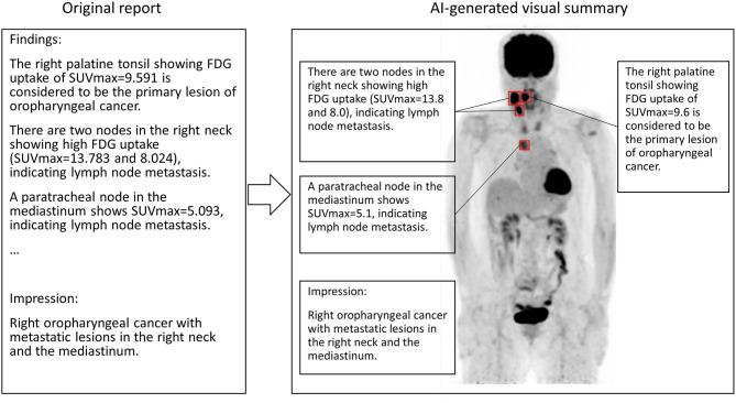
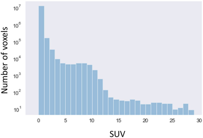
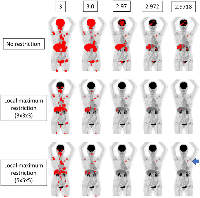
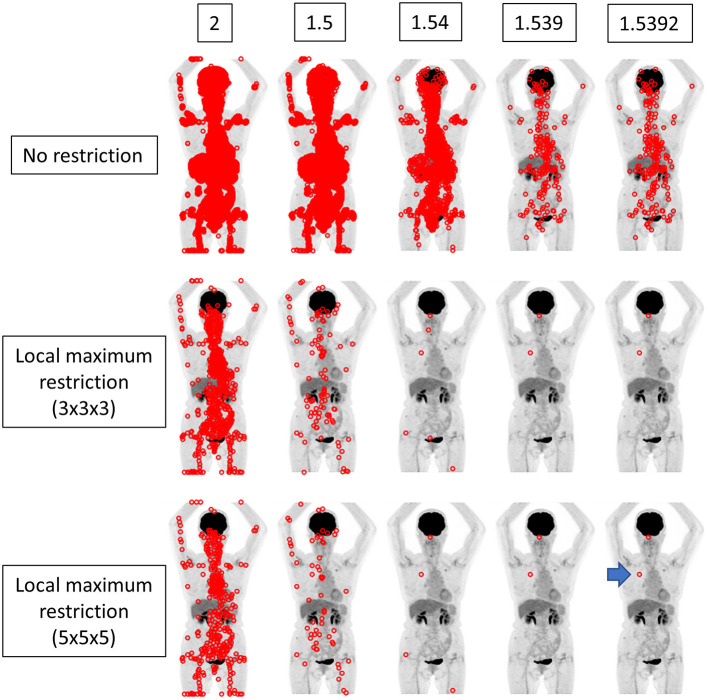
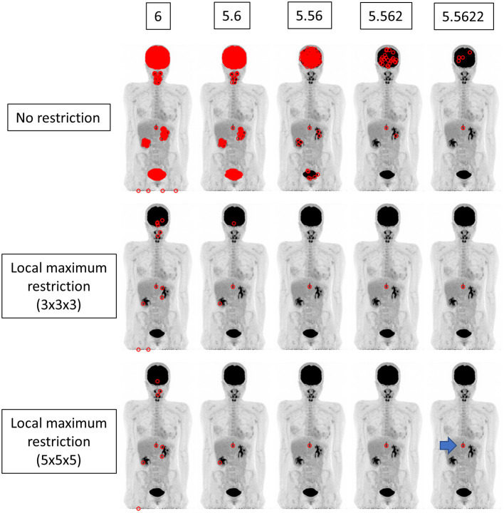
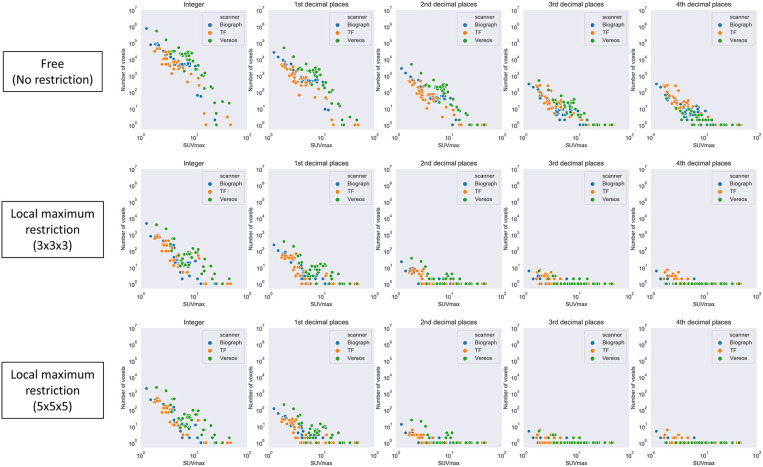
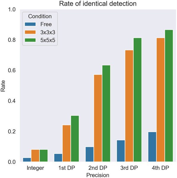
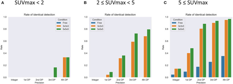
Diagnostic reports contribute not only to the particular patient, but also to constructing massive training dataset in the era of artificial intelligence (AI). The maximum standardized uptake value (SUVmax) is often described in daily diagnostic reports of [F] fluorodeoxyglucose (FDG) positron emission tomography (PET) - computed tomography (CT). If SUVmax can be used as an identifier of lesion, that would greatly help AI interpret diagnostic reports. We aimed to clarify whether the lesion can be localized using SUVmax strings. The institutional review board approved this retrospective study. We investigated a total of 112 lesions from 30 FDG PET-CT images acquired with 3 different scanners. SUVmax was calculated from DICOM files based on the latest Quantitative Imaging Biomarkers Alliance (QIBA) publication. The voxels showing the given SUVmax were exhaustively searched in the whole-body images and counted. SUVmax was provided with 5 different degrees of precision: integer (e.g., 3), 1st decimal places (DP) (3.1), 2nd DP (3.14), 3rd DP (3.142), and 4th DP (3.1416). For instance, when SUVmax = 3.14 was given, the voxels with 3.135 ≤ SUVmax < 3.145 were extracted. We also evaluated whether local maximum restriction could improve the identifying performance, where only the voxels showing the highest intensity within some neighborhood were considered. We defined that "identical detection" was achieved when only single voxel satisfied the criterion. A total of 112 lesions from 30 FDG PET-CT images were investigated. SUVmax ranged from 1.3 to 49.1 (median = 5.6). Generally, when larger and more precise SUVmax values were given, fewer voxels satisfied the criterion. The local maximum restriction was very effective. When SUVmax was determined to 4 decimal places (e.g., 3.1416) and the local maximum restriction was applied, identical detection was achieved in 33.3% (lesions with SUVmax < 2), 79.5% (2 ≤ SUVmax < 5), and 97.8% (5 ≤ SUVmax) of lesions. In this preliminary study, SUVmax of FDG PET-CT could be used as an identifier to localize the lesion if precise SUVmax is provided and local maximum restriction was applied, although the lesions showing SUVmax < 2 were difficult to identify. The proposed method may have potential to make use of diagnostic reports retrospectively for constructing training datasets for AI.

摘要

诊断报告不仅对特定患者有帮助,而且在人工智能(AI)时代有助于构建大规模训练数据集。最大标准化摄取值(SUVmax)常在[F]氟脱氧葡萄糖(FDG)正电子发射断层扫描(PET)-计算机断层扫描(CT)的日常诊断报告中描述。如果SUVmax可用作病变的标识符,将极大地帮助AI解读诊断报告。我们旨在阐明是否可以使用SUVmax字符串定位病变。机构审查委员会批准了这项回顾性研究。我们调查了用3种不同扫描仪获取的30张FDG PET-CT图像中的总共112个病变。基于最新的定量成像生物标志物联盟(QIBA)出版物从DICOM文件中计算SUVmax。在全身图像中详尽搜索显示给定SUVmax的体素并计数。SUVmax具有5种不同程度的精度:整数(例如,3)、保留1位小数(3.1)、保留2位小数(3.14)、保留3位小数(3.142)和保留4位小数(3.1416)。例如,当给出SUVmax = 3.14时,提取3.135≤SUVmax<3.145的体素。我们还评估了局部最大值限制是否可以提高识别性能,即仅考虑在某个邻域内显示最高强度的体素。我们定义当只有单个体素满足标准时实现“相同检测”。对30张FDG PET-CT图像中的总共112个病变进行了调查。SUVmax范围为1.3至49.1(中位数 = 5.6)。一般来说,当给出更大且更精确的SUVmax值时,满足标准的体素更少。局部最大值限制非常有效。当将SUVmax确定到4位小数(例如,3.1416)并应用局部最大值限制时,在SUVmax<2的病变中有33.3%、2≤SUVmax<5的病变中有79.5%以及5≤SUVmax的病变中有97.8%实现了相同检测。在这项初步研究中,如果提供精确的SUVmax并应用局部最大值限制,FDG PET-CT的SUVmax可以用作定位病变的标识符,尽管显示SUVmax<2的病变难以识别。所提出的方法可能有潜力回顾性地利用诊断报告来构建AI的训练数据集。

https://cdn.ncbi.nlm.nih.gov/pmc/blobs/0660/8113693/fde86c6f0062/fmed-08-647562-g0008.jpg
https://cdn.ncbi.nlm.nih.gov/pmc/blobs/0660/8113693/60484782c77c/fmed-08-647562-g0001.jpg
https://cdn.ncbi.nlm.nih.gov/pmc/blobs/0660/8113693/563624ace13e/fmed-08-647562-g0002.jpg
https://cdn.ncbi.nlm.nih.gov/pmc/blobs/0660/8113693/4a8eb610f3ab/fmed-08-647562-g0003.jpg
https://cdn.ncbi.nlm.nih.gov/pmc/blobs/0660/8113693/db2208056f1e/fmed-08-647562-g0004.jpg
https://cdn.ncbi.nlm.nih.gov/pmc/blobs/0660/8113693/fe040d3a88a3/fmed-08-647562-g0005.jpg
https://cdn.ncbi.nlm.nih.gov/pmc/blobs/0660/8113693/35bf6b0372d7/fmed-08-647562-g0006.jpg
https://cdn.ncbi.nlm.nih.gov/pmc/blobs/0660/8113693/38bf53502da0/fmed-08-647562-g0007.jpg
https://cdn.ncbi.nlm.nih.gov/pmc/blobs/0660/8113693/fde86c6f0062/fmed-08-647562-g0008.jpg

相似文献

1
A Preliminary Study to Use SUVmax of FDG PET-CT as an Identifier of Lesion for Artificial Intelligence.一项使用氟代脱氧葡萄糖正电子发射断层显像-计算机断层扫描(FDG PET-CT)的最大标准摄取值(SUVmax)作为人工智能病变识别指标的初步研究。
Front Med (Lausanne). 2021 Apr 28;8:647562. doi: 10.3389/fmed.2021.647562. eCollection 2021.
2
More advantages in detecting bone and soft tissue metastases from prostate cancer using F-PSMA PET/CT.使用F-PSMA PET/CT检测前列腺癌骨和软组织转移方面有更多优势。
Hell J Nucl Med. 2019 Jan-Apr;22(1):6-9. doi: 10.1967/s002449910952. Epub 2019 Mar 7.
3
Simultaneous whole body (18)F-fluorodeoxyglucose positron emission tomography magnetic resonance imaging for evaluation of pediatric cancer: Preliminary experience and comparison with (18)F-fluorodeoxyglucose positron emission tomography computed tomography.用于评估儿童癌症的同时全身(18)F-氟脱氧葡萄糖正电子发射断层扫描磁共振成像:初步经验及与(18)F-氟脱氧葡萄糖正电子发射断层扫描计算机断层扫描的比较
World J Radiol. 2016 Mar 28;8(3):322-30. doi: 10.4329/wjr.v8.i3.322.
4
(18)F-FPPRGD2 PET/CT: pilot phase evaluation of breast cancer patients.(18)F-FPPRGD2 PET/CT:乳腺癌患者的初步评估阶段。
Radiology. 2014 Nov;273(2):549-59. doi: 10.1148/radiol.14140028. Epub 2014 Jul 16.
5
Prognostic implication of fluorine-18 fluorodeoxyglucose positron emission tomography/computed tomography in patients with recurrent papillary thyroid cancer.氟-18氟脱氧葡萄糖正电子发射断层扫描/计算机断层扫描对复发性乳头状甲状腺癌患者的预后影响
Head Neck. 2018 Jan;40(1):94-102. doi: 10.1002/hed.24967. Epub 2017 Nov 11.
6
Improving the detection of small lesions using a state-of-the-art time-of-flight PET/CT system and small-voxel reconstructions.使用最先进的飞行时间PET/CT系统和小体素重建技术提高小病变的检测能力。
J Nucl Med Technol. 2015 Mar;43(1):21-7. doi: 10.2967/jnmt.114.147215. Epub 2015 Jan 22.
7
F-FDG PET/CT in diagnostic and prognostic evaluation of patients with cardiac masses: a retrospective study.F-FDG PET/CT在心脏肿块患者诊断和预后评估中的应用:一项回顾性研究
Eur J Nucl Med Mol Imaging. 2020 May;47(5):1083-1093. doi: 10.1007/s00259-019-04632-w. Epub 2019 Dec 5.
8
FDG PET/CT used in identifying adult-onset Still's disease in connective tissue diseases.18F-氟代脱氧葡萄糖正电子发射断层显像/计算机断层扫描在结缔组织病中用于识别成人斯蒂尔病。
Clin Rheumatol. 2020 Sep;39(9):2735-2742. doi: 10.1007/s10067-020-05041-3. Epub 2020 Mar 16.
9
Correlation Between F-FDG PET/CT Findings and BI-RADS Assessment Using Ultrasound in the Evaluation of Breast Lesions: A Multicenter Study.乳腺病变 F-FDG PET/CT 表现与超声 BI-RADS 评估的相关性:一项多中心研究。
Acad Radiol. 2020 May;27(5):682-688. doi: 10.1016/j.acra.2019.05.020. Epub 2019 Jul 13.
10
Diagnostic performance of F-FDG-PET-CT in adrenal lesions using histopathology as reference standard.F-FDG-PET-CT 对以组织病理学为参考标准的肾上腺病变的诊断性能。
Abdom Radiol (NY). 2017 Feb;42(2):577-584. doi: 10.1007/s00261-016-0915-4.

引用本文的文献

1
Correlation study of 18F-FDG PET/CT metabolic parameters, heterogeneity index, and microvascular invasion, and its nomogram potential in predicting microvascular invasion in liver cancer before liver transplantation.18F-FDG PET/CT代谢参数、异质性指数与微血管侵犯的相关性研究及其在预测肝癌肝移植术前微血管侵犯中的列线图潜力
Nucl Med Commun. 2025 Jun 24. doi: 10.1097/MNM.0000000000002014.
2
Correlation of preoperative 18F-FDG-PET/CT tumor staging and maximum standardized uptake values with preoperative CT, postoperative tumor classification, and histopathological parameters of oral squamous cell carcinoma.术前18F-FDG-PET/CT肿瘤分期及最大标准化摄取值与口腔鳞状细胞癌术前CT、术后肿瘤分级和组织病理学参数的相关性
Clin Oral Investig. 2025 Mar 18;29(4):189. doi: 10.1007/s00784-025-06252-1.
3

本文引用的文献

1
The implementation of natural language processing to extract index lesions from breast magnetic resonance imaging reports.运用自然语言处理技术从乳腺磁共振成像报告中提取病灶索引。
BMC Med Inform Decis Mak. 2019 Dec 30;19(1):288. doi: 10.1186/s12911-019-0997-3.
2
Finding relevant free-text radiology reports at scale with IBM Watson Content Analytics: a feasibility study in the UK NHS.使用IBM Watson内容分析大规模查找相关的自由文本放射学报告:英国国民健康服务体系中的一项可行性研究
J Biomed Semantics. 2019 Nov 12;10(Suppl 1):21. doi: 10.1186/s13326-019-0213-5.
3
Natural Language Processing Approaches to Detect the Timeline of Metastatic Recurrence of Breast Cancer.
Early prediction of microvascular invasion (MVI) occurrence in hepatocellular carcinoma (HCC) by F-FDG PET/CT and laboratory data.基于 F-FDG PET/CT 和实验室数据对肝细胞癌(HCC)中微血管侵犯(MVI)发生的早期预测。
Eur J Med Res. 2024 Jul 30;29(1):395. doi: 10.1186/s40001-024-01973-7.
4
Diagnostics of atherosclerosis: Overview of the existing methods.动脉粥样硬化的诊断:现有方法概述。
Front Cardiovasc Med. 2023 May 9;10:1134097. doi: 10.3389/fcvm.2023.1134097. eCollection 2023.
5
Medical Radiation Exposure Reduction in PET via Super-Resolution Deep Learning Model.通过超分辨率深度学习模型减少正电子发射断层扫描中的医学辐射暴露
Diagnostics (Basel). 2022 Mar 31;12(4):872. doi: 10.3390/diagnostics12040872.
6
Tumor Size Measurements for Predicting Hodgkin's and Non-Hodgkin's Lymphoma Response to Treatment.用于预测霍奇金淋巴瘤和非霍奇金淋巴瘤治疗反应的肿瘤大小测量
Metabolites. 2022 Mar 24;12(4):285. doi: 10.3390/metabo12040285.
7
Artificial Intelligence in Lymphoma PET Imaging:: A Scoping Review (Current Trends and Future Directions).人工智能在淋巴瘤 PET 成像中的应用:范围综述(当前趋势和未来方向)。
PET Clin. 2022 Jan;17(1):145-174. doi: 10.1016/j.cpet.2021.09.006.
用于检测乳腺癌转移复发时间线的自然语言处理方法
JCO Clin Cancer Inform. 2019 Oct;3:1-12. doi: 10.1200/CCI.19.00034.
4
Deep learning to convert unstructured CT pulmonary angiography reports into structured reports.深度学习将非结构化CT肺血管造影报告转换为结构化报告。
Eur Radiol Exp. 2019 Sep 23;3(1):37. doi: 10.1186/s41747-019-0118-1.
5
Determining Follow-Up Imaging Study Using Radiology Reports.基于放射科报告判断是否需要进行随访影像学检查。
J Digit Imaging. 2020 Feb;33(1):121-130. doi: 10.1007/s10278-019-00260-w.
6
Nuclear medicine practice in Japan: a report of the eighth nationwide survey in 2017.日本核医学实践状况:2017 年第八次全国调查结果报告
Ann Nucl Med. 2019 Oct;33(10):725-732. doi: 10.1007/s12149-019-01382-5. Epub 2019 Jun 24.
7
Endorsement of International Consensus Radiochemistry Nomenclature Guidelines.国际放射化学命名法共识指南认可
Eur J Nucl Med Mol Imaging. 2019 Jul;46(7):1399. doi: 10.1007/s00259-018-4243-5. Epub 2019 Apr 16.
8
Volume-based parameters on FDG PET may predict the proliferative potential of soft-tissue sarcomas.基于体素的FDG PET参数可能预测软组织肉瘤的增殖潜能。
Ann Nucl Med. 2019 Jan;33(1):22-31. doi: 10.1007/s12149-018-1298-0. Epub 2018 Sep 8.
9
Reproducibility and uptake time dependency of volume-based parameters on FDG-PET for lung cancer.基于体积的参数在肺癌氟代脱氧葡萄糖正电子发射断层扫描(FDG-PET)中的可重复性及摄取时间依赖性
BMC Cancer. 2016 Aug 2;16:576. doi: 10.1186/s12885-016-2624-3.
10
Image Quality and Diagnostic Performance of a Digital PET Prototype in Patients with Oncologic Diseases: Initial Experience and Comparison with Analog PET.数字正电子发射断层显像(PET)原型机在肿瘤疾病患者中的图像质量与诊断性能:初步经验及与模拟PET的比较
J Nucl Med. 2015 Sep;56(9):1378-85. doi: 10.2967/jnumed.114.148338. Epub 2015 Jul 9.