Lee Emerson E, Andresen Nicholas S, McKenzie Bryan, Sharon Jeffrey D, Francis Howard W, Sun Daniel Q
Department of Otolaryngology-Head and Neck Surgery, Johns Hopkins University, USA.
Inova Fairfax Hospital, USA.
World J Otorhinolaryngol Head Neck Surg. 2021 Jan 24;7(2):71-81. doi: 10.1016/j.wjorl.2020.12.005. eCollection 2021 Apr.
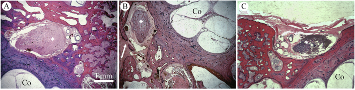
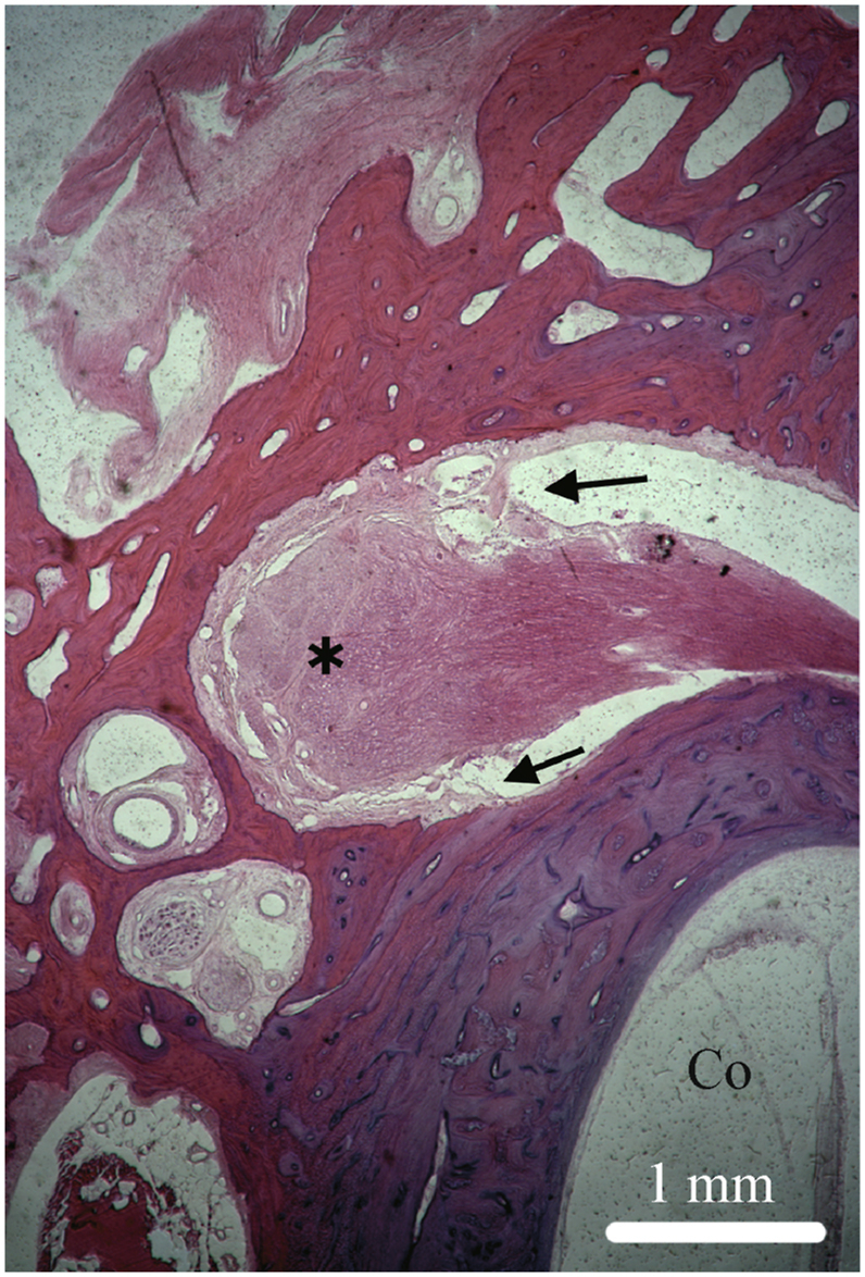
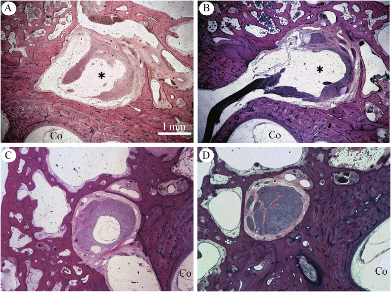
Cerebrospinal fluid (CSF) fistulae originating from the fallopian canal of the facial nerve is hypothesized to arise due to atypical patterns of subarachnoid space extension into the geniculate ganglion or more distal regions along the intratemporal course of the facial nerve, but its pathogenesis remains poorly understood. Although a rare etiology of CSF fistulae of the temporal bone, there are significant clinical ramifications due to the risk of recurrent meningitis, difficulty in identifying the anatomic location of the CSF leak, and technical challenges associated with surgical repair. We present three clinical cases of arachnoid cysts within the geniculate fossa with or without CSF fistulization and provide histopathologic correlates of this rare clinical phenomenon from a human temporal bone collection. The pediatric and adult patients presented suggest differential pathophysiologic mechanisms associated with CSF fistulae. Temporal bone histology reveals atypical patterns of subarachnoid space extension in the fallopian canal that may underlie arachnoid cyst formation and overt CSF leak from the geniculate region.
源自面神经鼓室段的脑脊液(CSF)瘘被推测是由于蛛网膜下腔向膝状神经节或沿面神经颞内段更远端区域延伸的非典型模式所致,但其发病机制仍知之甚少。尽管颞骨CSF瘘的病因罕见,但由于存在复发性脑膜炎的风险、难以确定CSF漏的解剖位置以及与手术修复相关的技术挑战,因此具有重大的临床影响。我们展示了三例膝状窝内伴有或不伴有CSF瘘形成的蛛网膜囊肿临床病例,并从人类颞骨标本中提供了这种罕见临床现象的组织病理学关联。所呈现的儿科和成人患者提示了与CSF瘘相关的不同病理生理机制。颞骨组织学显示鼓室内蛛网膜下腔延伸的非典型模式,这可能是蛛网膜囊肿形成以及膝状区域明显CSF漏的基础。