South West Thames Institute for Renal Research, London, SM5 1AA, U.K.
St Georges' University of London, SW17 0RE, U.K.
Biosci Rep. 2021 Jun 25;41(6). doi: 10.1042/BSR20203947.
In the kidney glucose is freely filtered by the glomerulus and, mainly, reabsorbed by sodium glucose cotransporter 2 (SGLT2) expressed in the early proximal tubule. Human proximal tubule epithelial cells (PTECs) undergo pathological and fibrotic changes seen in diabetic kidney disease (DKD) in response to elevated glucose. We developed a specific in vitro model of DKD using primary human PTECs with exposure to high D-glucose and TGF-β1 and propose a role for SGLT2 inhibition in regulating fibrosis.
Western blotting was performed to detect cellular and secreted proteins as well as phosphorylated intracellular signalling proteins. qPCR was used to detect CCN2 RNA. Gamma glutamyl transferase (GT) activity staining was performed to confirm PTEC phenotype. SGLT2 and ERK inhibition on high D-glucose, 25 mM, and TGF-β1, 0.75 ng/ml, treated cells was explored using dapagliflozin and U0126, respectively.
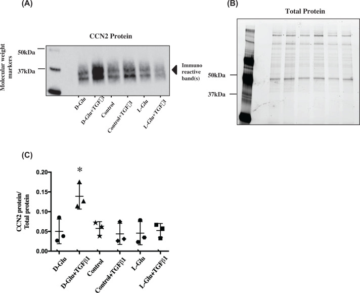
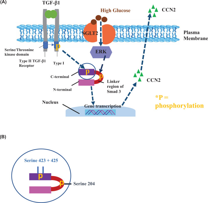
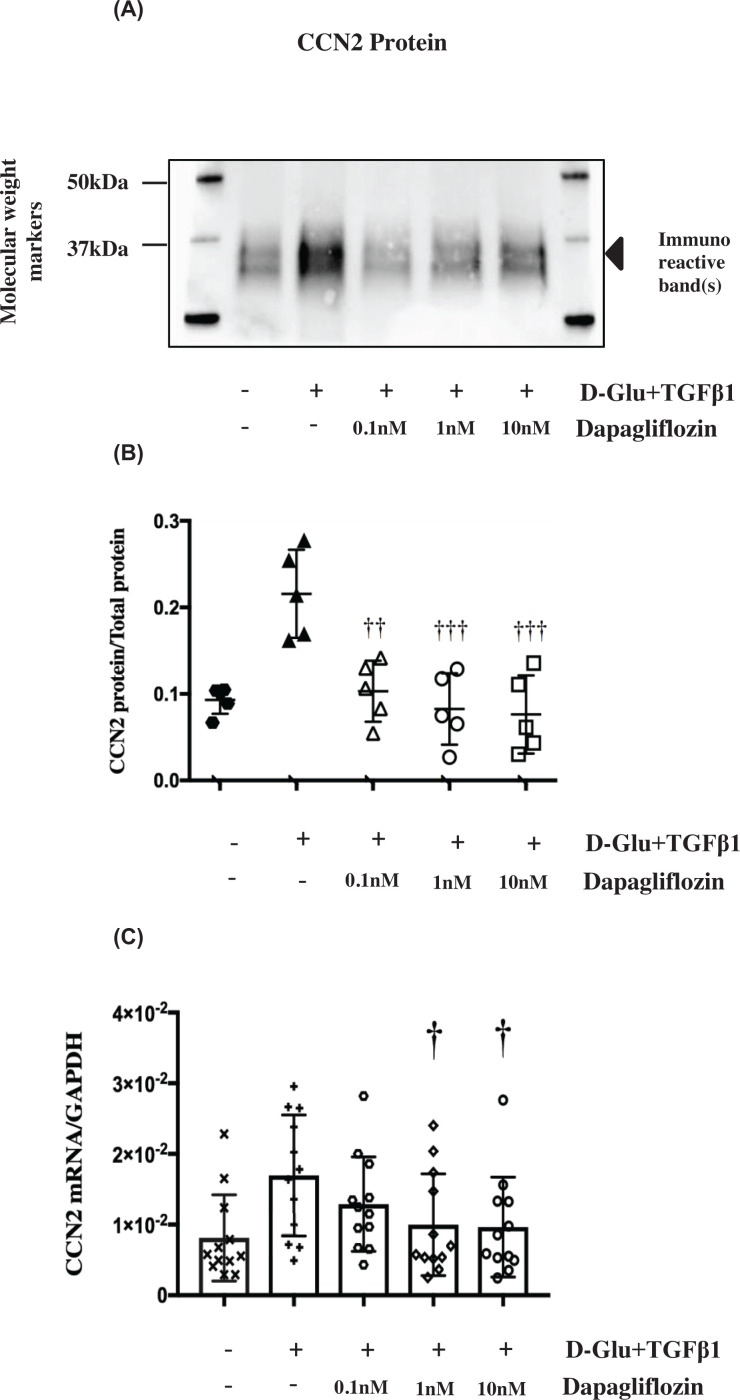
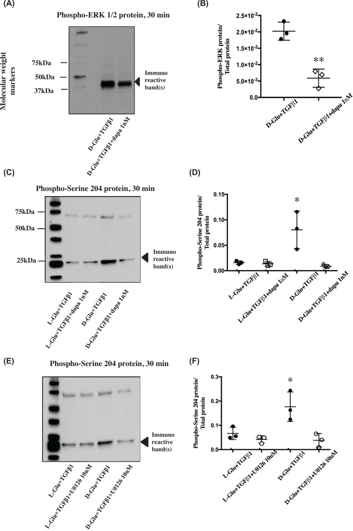
Only the combination of high D-glucose and TGF-β1 treatment significantly up-regulated CCN2 RNA and protein expression. This increase was significantly ameliorated by dapagliflozin. High D-glucose treatment raised phospho ERK which was also inhibited by dapagliflozin. TGF-β1 increased cellular phospho SSXS Smad3 serine 423 and 425, with and without high D-glucose. Glucose alone had no effect. Smad3 serine 204 phosphorylation was significantly raised by a combination of high D-glucose+TGF-β1; this rise was significantly reduced by both SGLT2 and MEK inhibition.
We show that high D-glucose and TGF-β1 are both required for CCN2 expression. This treatment also caused Smad3 linker region phosphorylation. Both outcomes were inhibited by dapagliflozin. We have identified a novel SGLT2 -ERK mediated promotion of TGF-β1/Smad3 signalling inducing a pro-fibrotic growth factor secretion. Our data evince support for substantial renoprotective benefits of SGLT2 inhibition in the diabetic kidney.
在肾脏中,葡萄糖可自由地被肾小球滤过,并且主要通过在近端肾小管早期表达的钠-葡萄糖协同转运蛋白 2(SGLT2)被重吸收。人近端肾小管上皮细胞(PTEC)在高葡萄糖作用下会发生糖尿病肾病(DKD)中所见的病理和纤维化改变。我们使用暴露于高 D-葡萄糖和 TGF-β1 的原代人 PTEC 开发了一种特定的 DKD 体外模型,并提出 SGLT2 抑制在调节纤维化中的作用。
通过 Western blot 检测细胞和分泌蛋白以及磷酸化细胞内信号蛋白,通过 qPCR 检测 CCN2 RNA。谷氨酰转移酶(GT)活性染色用于确认 PTEC 表型。使用 dapagliflozin 和 U0126 分别研究 SGLT2 和 ERK 在高 D-葡萄糖(25 mM)和 TGF-β1(0.75 ng/ml)处理细胞中的抑制作用。
仅高 D-葡萄糖和 TGF-β1 联合处理可显著上调 CCN2 RNA 和蛋白表达。 dapagliflozin 可显著改善这种增加。高 D-葡萄糖处理可提高磷酸化 ERK,dapagliflozin 也可抑制磷酸化 ERK。TGF-β1 可增加细胞内磷酸化 SSXS Smad3 丝氨酸 423 和 425,无论是否存在高 D-葡萄糖。单独的葡萄糖没有作用。高 D-葡萄糖+TGF-β1 联合处理可显著提高 Smad3 丝氨酸 204 磷酸化;SGLT2 和 MEK 抑制均可显著降低此升高。
我们表明高 D-葡萄糖和 TGF-β1 均是 CCN2 表达所必需的。这种处理还引起 Smad3 连接区磷酸化。两种结果均被 dapagliflozin 抑制。我们已经确定了一种新的 SGLT2-ERK 介导的 TGF-β1/Smad3 信号转导促进促纤维化生长因子分泌。我们的数据支持 SGLT2 抑制在糖尿病肾脏中具有实质性的肾脏保护作用。