• 文献检索
  • 文档翻译
  • 深度研究
  • 学术资讯
  • Suppr Zotero 插件Zotero 插件
  • 邀请有礼
  • 套餐&价格
  • 历史记录
应用&插件
Suppr Zotero 插件Zotero 插件浏览器插件Mac 客户端Windows 客户端微信小程序
定价
高级版会员购买积分包购买API积分包
服务
文献检索文档翻译深度研究API 文档MCP 服务
关于我们
关于 Suppr公司介绍联系我们用户协议隐私条款
关注我们

Suppr 超能文献

核心技术专利:CN118964589B侵权必究
粤ICP备2023148730 号-1Suppr @ 2026

文献检索

告别复杂PubMed语法,用中文像聊天一样搜索,搜遍4000万医学文献。AI智能推荐,让科研检索更轻松。

立即免费搜索

文件翻译

保留排版,准确专业,支持PDF/Word/PPT等文件格式,支持 12+语言互译。

免费翻译文档

深度研究

AI帮你快速写综述,25分钟生成高质量综述,智能提取关键信息,辅助科研写作。

立即免费体验

单细胞转录组分析定义了小鼠胰腺导管树的异质性。

Single-cell transcriptome analysis defines heterogeneity of the murine pancreatic ductal tree.

机构信息

Diabetes Center, University of California, San Francisco, San Francisco, United States.

Center for Bioengineering and Tissue Regeneration, University of California, San Francisco, San Francisco, United States.

出版信息

Elife. 2021 May 19;10:e67776. doi: 10.7554/eLife.67776.

DOI:10.7554/eLife.67776
PMID:34009124
原文链接:https://pmc.ncbi.nlm.nih.gov/articles/PMC8184217/
Abstract

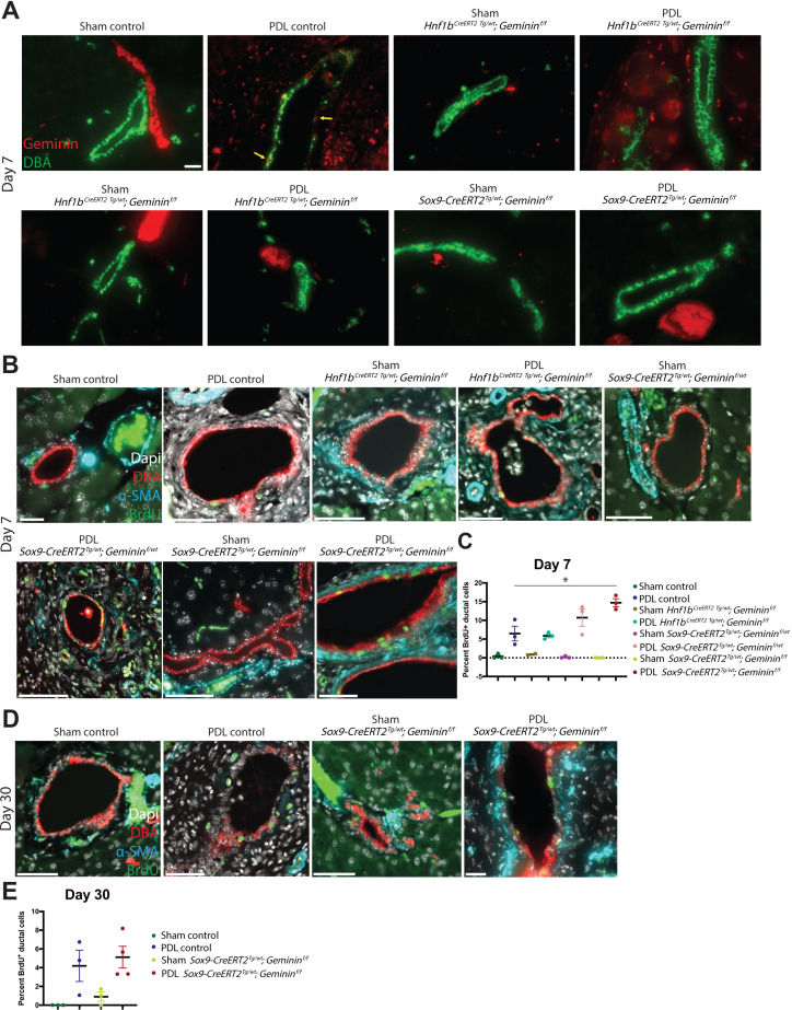
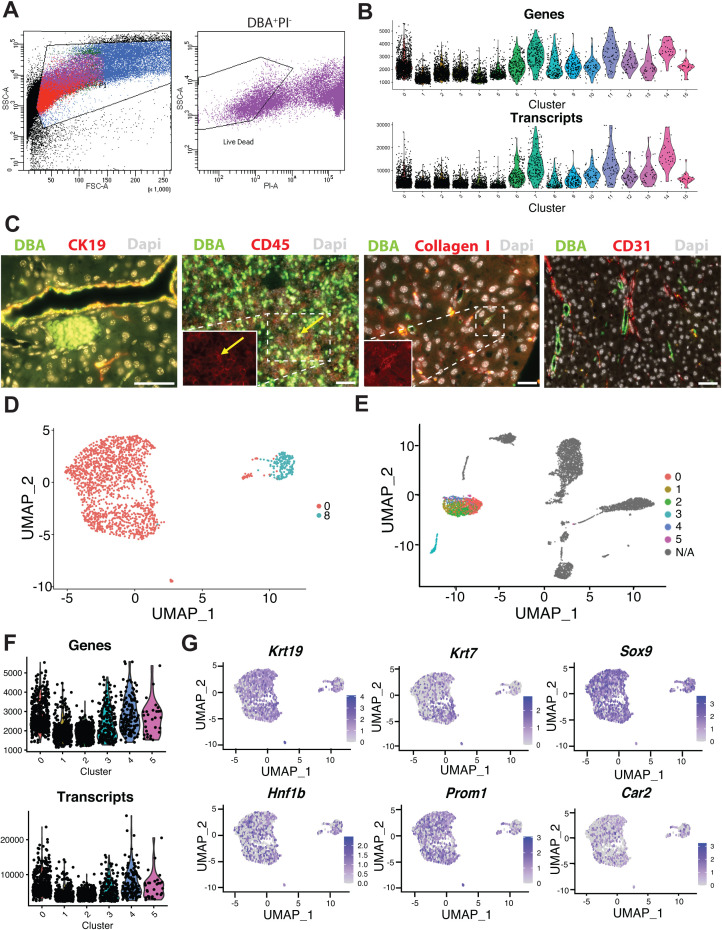
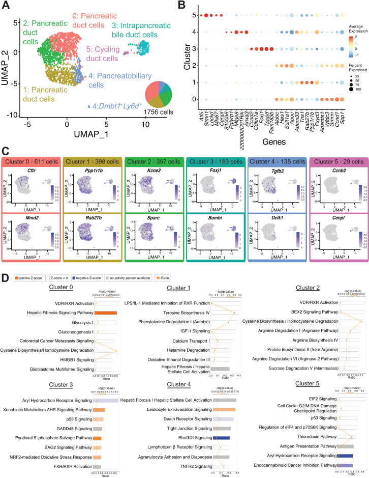
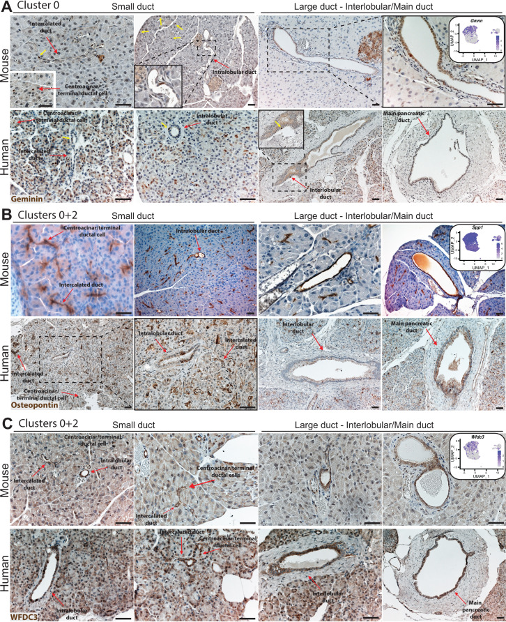
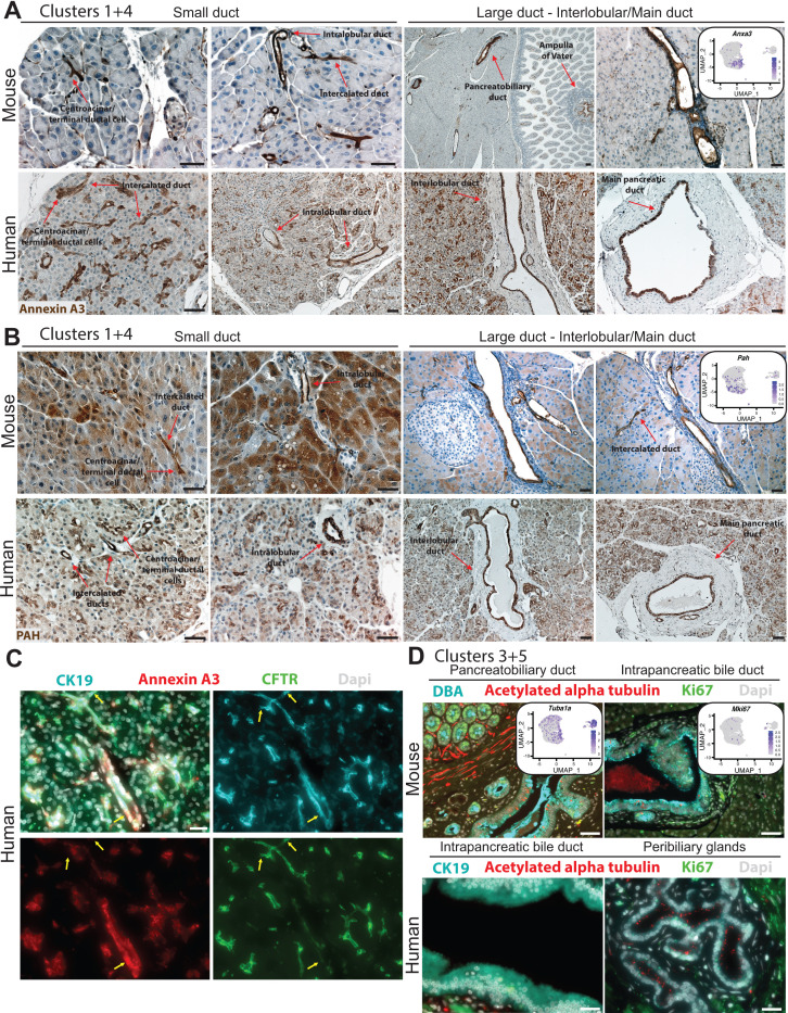
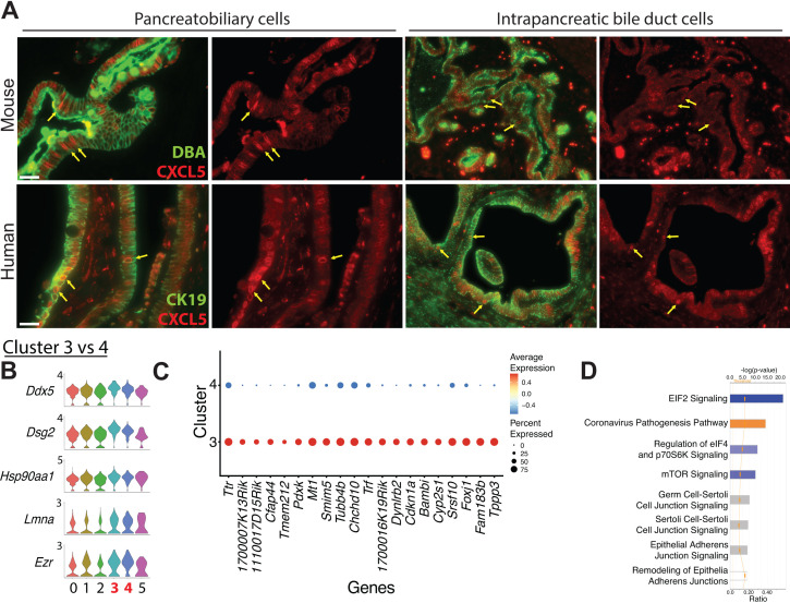
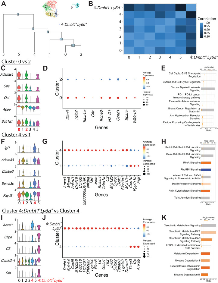
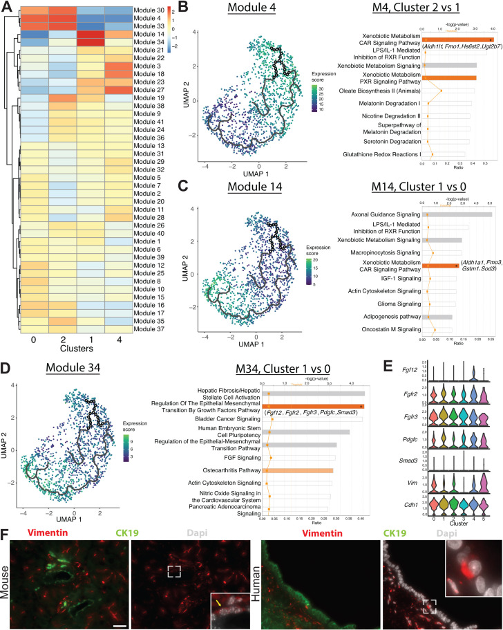
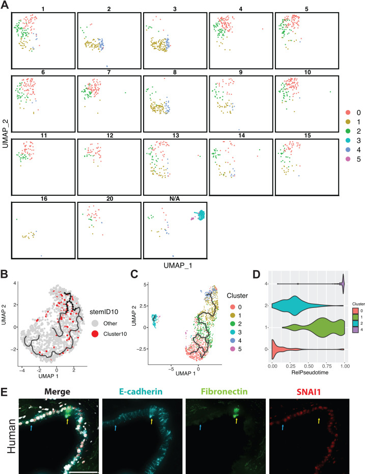
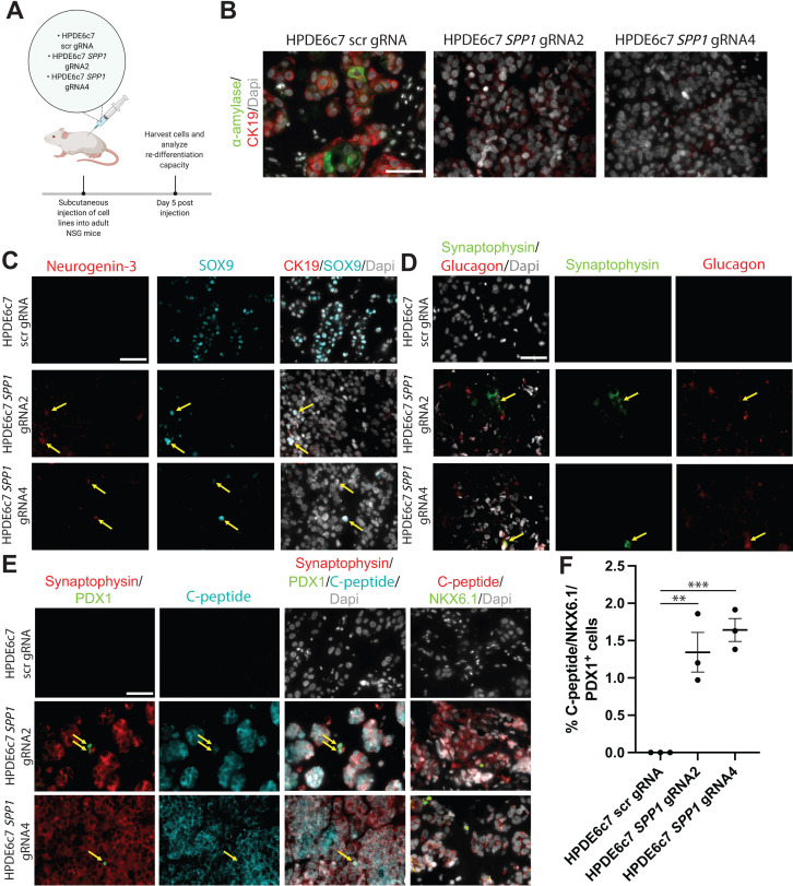
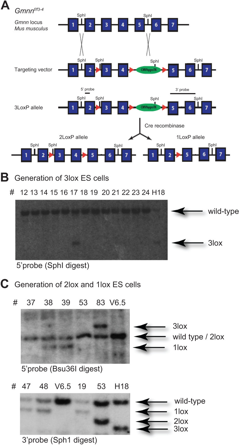
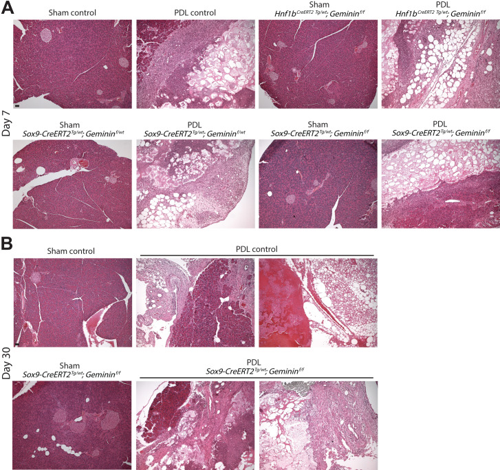
To study disease development, an inventory of an organ's cell types and understanding of physiologic function is paramount. Here, we performed single-cell RNA-sequencing to examine heterogeneity of murine pancreatic duct cells, pancreatobiliary cells, and intrapancreatic bile duct cells. We describe an epithelial-mesenchymal transitory axis in our three pancreatic duct subpopulations and identify osteopontin as a regulator of this fate decision as well as human duct cell dedifferentiation. Our results further identify functional heterogeneity within pancreatic duct subpopulations by elucidating a role for geminin in accumulation of DNA damage in the setting of chronic pancreatitis. Our findings implicate diverse functional roles for subpopulations of pancreatic duct cells in maintenance of duct cell identity and disease progression and establish a comprehensive road map of murine pancreatic duct cell, pancreatobiliary cell, and intrapancreatic bile duct cell homeostasis.

摘要

为了研究疾病的发展,了解器官的细胞类型和生理功能是至关重要的。在这里,我们进行了单细胞 RNA 测序,以研究小鼠胰腺导管细胞、胰胆管细胞和胰内胆管细胞的异质性。我们描述了我们的三个胰腺导管亚群中的上皮-间充质过渡轴,并确定骨桥蛋白是这种命运决定以及人胆管细胞去分化的调节剂。我们的研究结果还通过阐明在慢性胰腺炎中 geminin 在积累 DNA 损伤中的作用,进一步阐明了胰腺导管亚群内的功能异质性。我们的发现表明,胰腺导管细胞亚群在维持导管细胞特征和疾病进展方面具有不同的功能作用,并建立了小鼠胰腺导管细胞、胰胆管细胞和胰内胆管细胞稳态的综合路线图。

https://cdn.ncbi.nlm.nih.gov/pmc/blobs/b6b7/8184217/9d5e728215c5/elife-67776-fig8-figsupp3.jpg
https://cdn.ncbi.nlm.nih.gov/pmc/blobs/b6b7/8184217/e3d1fa4105fd/elife-67776-fig1.jpg
https://cdn.ncbi.nlm.nih.gov/pmc/blobs/b6b7/8184217/f0dca17cad64/elife-67776-fig1-figsupp1.jpg
https://cdn.ncbi.nlm.nih.gov/pmc/blobs/b6b7/8184217/733b1fc8d657/elife-67776-fig2.jpg
https://cdn.ncbi.nlm.nih.gov/pmc/blobs/b6b7/8184217/be0ca37f7669/elife-67776-fig2-figsupp1.jpg
https://cdn.ncbi.nlm.nih.gov/pmc/blobs/b6b7/8184217/f4c4a09f7902/elife-67776-fig2-figsupp2.jpg
https://cdn.ncbi.nlm.nih.gov/pmc/blobs/b6b7/8184217/3c4cd729ff26/elife-67776-fig2-figsupp3.jpg
https://cdn.ncbi.nlm.nih.gov/pmc/blobs/b6b7/8184217/9a1c8343003c/elife-67776-fig10.jpg
https://cdn.ncbi.nlm.nih.gov/pmc/blobs/b6b7/8184217/42734655d7d2/elife-67776-fig3.jpg
https://cdn.ncbi.nlm.nih.gov/pmc/blobs/b6b7/8184217/20bb10085f4f/elife-67776-fig3-figsupp1.jpg
https://cdn.ncbi.nlm.nih.gov/pmc/blobs/b6b7/8184217/e134eb436f5f/elife-67776-fig3-figsupp2.jpg
https://cdn.ncbi.nlm.nih.gov/pmc/blobs/b6b7/8184217/03638571840b/elife-67776-fig3-figsupp3.jpg
https://cdn.ncbi.nlm.nih.gov/pmc/blobs/b6b7/8184217/175b0115b090/elife-67776-fig4.jpg
https://cdn.ncbi.nlm.nih.gov/pmc/blobs/b6b7/8184217/a9a0dee68891/elife-67776-fig5.jpg
https://cdn.ncbi.nlm.nih.gov/pmc/blobs/b6b7/8184217/6aad4ebebc69/elife-67776-fig5-figsupp1.jpg
https://cdn.ncbi.nlm.nih.gov/pmc/blobs/b6b7/8184217/b29a345dbf40/elife-67776-fig6.jpg
https://cdn.ncbi.nlm.nih.gov/pmc/blobs/b6b7/8184217/1bc7b9407e2e/elife-67776-fig6-figsupp1.jpg
https://cdn.ncbi.nlm.nih.gov/pmc/blobs/b6b7/8184217/09de1b8f0e4c/elife-67776-fig7.jpg
https://cdn.ncbi.nlm.nih.gov/pmc/blobs/b6b7/8184217/8ab9bc1e8fcd/elife-67776-fig8.jpg
https://cdn.ncbi.nlm.nih.gov/pmc/blobs/b6b7/8184217/562dc2fd7141/elife-67776-fig8-figsupp1.jpg
https://cdn.ncbi.nlm.nih.gov/pmc/blobs/b6b7/8184217/02dac3099502/elife-67776-fig8-figsupp2.jpg
https://cdn.ncbi.nlm.nih.gov/pmc/blobs/b6b7/8184217/9d5e728215c5/elife-67776-fig8-figsupp3.jpg

相似文献

1
Single-cell transcriptome analysis defines heterogeneity of the murine pancreatic ductal tree.单细胞转录组分析定义了小鼠胰腺导管树的异质性。
Elife. 2021 May 19;10:e67776. doi: 10.7554/eLife.67776.
2
A Single-Cell Atlas of the Murine Pancreatic Ductal Tree Identifies Novel Cell Populations With Potential Implications in Pancreas Regeneration and Exocrine Pathogenesis.单细胞图谱描绘了小鼠胰腺导管树,鉴定了具有潜在胰腺再生和外分泌发病机制意义的新细胞群体。
Gastroenterology. 2024 Oct;167(5):944-960.e15. doi: 10.1053/j.gastro.2024.06.008. Epub 2024 Jun 21.
3
The Crosstalk between Nrf2 and TGF-β1 in the Epithelial-Mesenchymal Transition of Pancreatic Duct Epithelial Cells.Nrf2与TGF-β1在胰腺导管上皮细胞上皮-间质转化中的相互作用
PLoS One. 2015 Jul 30;10(7):e0132978. doi: 10.1371/journal.pone.0132978. eCollection 2015.
4
Pancreatic duct glands are distinct ductal compartments that react to chronic injury and mediate Shh-induced metaplasia.胰腺导管腺是独特的导管隔室,对慢性损伤有反应,并介导 Shh 诱导的化生。
Gastroenterology. 2010 Mar;138(3):1166-77. doi: 10.1053/j.gastro.2009.12.005. Epub 2009 Dec 21.
5
Complement Component 5 Mediates Development of Fibrosis, via Activation of Stellate Cells, in 2 Mouse Models of Chronic Pancreatitis.在2种慢性胰腺炎小鼠模型中,补体成分5通过激活星状细胞介导纤维化的发展。
Gastroenterology. 2015 Sep;149(3):765-76.e10. doi: 10.1053/j.gastro.2015.05.012. Epub 2015 May 19.
6
Identification and manipulation of biliary metaplasia in pancreatic tumors.鉴定和操控胰腺肿瘤中的胆管化生。
Gastroenterology. 2014 Jan;146(1):233-44.e5. doi: 10.1053/j.gastro.2013.08.053. Epub 2013 Aug 30.
7
Single-cell-resolved differentiation of human induced pluripotent stem cells into pancreatic duct-like organoids on a microwell chip.在微井芯片上,单细胞解析的人诱导多能干细胞向胰腺导管样类器官的分化。
Nat Biomed Eng. 2021 Aug;5(8):897-913. doi: 10.1038/s41551-021-00757-2. Epub 2021 Jul 8.
8
Novel Approach for Pancreas Transcriptomics Reveals the Cellular Landscape in Homeostasis and Acute Pancreatitis.胰腺转录组学的新方法揭示了在稳态和急性胰腺炎中的细胞景观。
Gastroenterology. 2024 Jun;166(6):1100-1113. doi: 10.1053/j.gastro.2024.01.043. Epub 2024 Feb 6.
9
Osteopontin is a novel marker of pancreatic ductal tissues and of undifferentiated pancreatic precursors in mice.骨桥蛋白是小鼠胰腺导管组织和未分化胰腺前体的一种新型标志物。
Dev Dyn. 2006 Jun;235(6):1659-67. doi: 10.1002/dvdy.20729.
10
Single-cell analyses of human pancreas: characteristics of two populations of acinar cells in chronic pancreatitis.单细胞分析人类胰腺:慢性胰腺炎中两种胰岛细胞的特征。
Am J Physiol Gastrointest Liver Physiol. 2021 Nov 1;321(5):G449-G460. doi: 10.1152/ajpgi.00482.2020. Epub 2021 Sep 15.

引用本文的文献

1
Pancreatic β-cell regeneration in situ by the ALK3 agonist THR-123.ALK3激动剂THR-123介导的胰腺β细胞原位再生
Nat Commun. 2025 Jul 3;16(1):6121. doi: 10.1038/s41467-025-61534-2.
2
Small molecules enhance the high-efficiency generation of pancreatic ductal organoids.小分子增强胰腺导管类器官的高效生成。
Acta Biochim Biophys Sin (Shanghai). 2024 Dec 5;57(7):1184-1194. doi: 10.3724/abbs.2024218.
3
Identification of mouse and human embryonic pancreatic cells with adult Procr progenitor transcriptomic and epigenomic characteristics.

本文引用的文献

1
Loss of HIF1A From Pancreatic Cancer Cells Increases Expression of PPP1R1B and Degradation of p53 to Promote Invasion and Metastasis.胰腺癌细胞中 HIF1A 的缺失会增加 PPP1R1B 的表达并降解 p53,从而促进侵袭和转移。
Gastroenterology. 2020 Nov;159(5):1882-1897.e5. doi: 10.1053/j.gastro.2020.07.046. Epub 2020 Aug 5.
2
microRNA-382 suppresses the progression of pancreatic cancer through the PI3K/Akt signaling pathway by inhibition of Anxa3.miRNA-382 通过抑制 Anxa3 抑制 PI3K/Akt 信号通路抑制胰腺癌的进展。
Am J Physiol Gastrointest Liver Physiol. 2020 Sep 1;319(3):G309-G322. doi: 10.1152/ajpgi.00322.2019. Epub 2020 May 28.
3
鉴定具有成年Procr祖细胞转录组和表观基因组特征的小鼠和人类胚胎胰腺细胞。
Front Endocrinol (Lausanne). 2025 Feb 13;16:1543960. doi: 10.3389/fendo.2025.1543960. eCollection 2025.
4
Exploring human pancreatic organoid modelling through single-cell RNA sequencing analysis.通过单细胞 RNA 测序分析探索人类胰腺类器官建模。
Commun Biol. 2024 Nov 18;7(1):1527. doi: 10.1038/s42003-024-07193-3.
5
Lineage tracing of stem cell-derived dopamine grafts in a Parkinson's model reveals shared origin of all graft-derived cells.在帕金森病模型中,对干细胞源性多巴胺移植的谱系追踪揭示了所有移植源性细胞的共同起源。
Sci Adv. 2024 Oct 18;10(42):eadn3057. doi: 10.1126/sciadv.adn3057.
6
Comparative transcriptomic analysis reveals the molecular changes of acute pancreatitis in experimental models.比较转录组学分析揭示了实验模型中急性胰腺炎的分子变化。
World J Gastroenterol. 2024 Apr 14;30(14):2038-2058. doi: 10.3748/wjg.v30.i14.2038.
7
Novel Approach for Pancreas Transcriptomics Reveals the Cellular Landscape in Homeostasis and Acute Pancreatitis.胰腺转录组学的新方法揭示了在稳态和急性胰腺炎中的细胞景观。
Gastroenterology. 2024 Jun;166(6):1100-1113. doi: 10.1053/j.gastro.2024.01.043. Epub 2024 Feb 6.
8
Oncogenic dependency plays a dominant role in the immune response to cancer.致癌依赖性在癌症的免疫反应中起着主导作用。
Proc Natl Acad Sci U S A. 2023 Oct 10;120(41):e2308635120. doi: 10.1073/pnas.2308635120. Epub 2023 Oct 2.
9
New discoveries in the field of metabolism by applying single-cell and spatial omics.通过应用单细胞和空间组学在代谢领域取得的新发现。
J Pharm Anal. 2023 Jul;13(7):711-725. doi: 10.1016/j.jpha.2023.06.002. Epub 2023 Jun 4.
10
Rediscovering Primary Cilia in Pancreatic Islets.重新发现胰腺胰岛中的初级纤毛。
Diabetes Metab J. 2023 Jul;47(4):454-469. doi: 10.4093/dmj.2022.0442. Epub 2023 Apr 28.
Single-cell resolution analysis of the human pancreatic ductal progenitor cell niche.
人类胰腺导管祖细胞生态位的单细胞分辨率分析。
Proc Natl Acad Sci U S A. 2020 May 19;117(20):10876-10887. doi: 10.1073/pnas.1918314117. Epub 2020 Apr 30.
4
Defining the emergence of myeloid-derived suppressor cells in breast cancer using single-cell transcriptomics.利用单细胞转录组学定义乳腺癌中髓系来源抑制细胞的出现。
Sci Immunol. 2020 Feb 21;5(44). doi: 10.1126/sciimmunol.aay6017.
5
Aldh1b1 expression defines progenitor cells in the adult pancreas and is required for Kras-induced pancreatic cancer.Aldh1b1 表达定义了成年胰腺中的祖细胞,并且是 Kras 诱导的胰腺肿瘤所必需的。
Proc Natl Acad Sci U S A. 2019 Oct 8;116(41):20679-20688. doi: 10.1073/pnas.1901075116. Epub 2019 Sep 23.
6
Transcriptional control of subtype switching ensures adaptation and growth of pancreatic cancer.转录控制亚型转换确保了胰腺癌的适应和生长。
Elife. 2019 May 28;8:e45313. doi: 10.7554/eLife.45313.
7
Single-Cell Analysis of the Liver Epithelium Reveals Dynamic Heterogeneity and an Essential Role for YAP in Homeostasis and Regeneration.单细胞分析肝脏上皮细胞揭示了动态异质性和 YAP 在稳态和再生中的重要作用。
Cell Stem Cell. 2019 Jul 3;25(1):23-38.e8. doi: 10.1016/j.stem.2019.04.004. Epub 2019 May 9.
8
Scrublet: Computational Identification of Cell Doublets in Single-Cell Transcriptomic Data.Scrublet:单细胞转录组数据中细胞二聚体的计算鉴定。
Cell Syst. 2019 Apr 24;8(4):281-291.e9. doi: 10.1016/j.cels.2018.11.005. Epub 2019 Apr 3.
9
The single-cell transcriptional landscape of mammalian organogenesis.哺乳动物器官发生的单细胞转录组图谱。
Nature. 2019 Feb;566(7745):496-502. doi: 10.1038/s41586-019-0969-x. Epub 2019 Feb 20.
10
Lineage dynamics of murine pancreatic development at single-cell resolution.单细胞分辨率解析的小鼠胰腺发育的谱系动力学。
Nat Commun. 2018 Sep 25;9(1):3922. doi: 10.1038/s41467-018-06176-3.