Nestle Ursula, Le Pechoux Cecile, De Ruysscher Dirk
Department of Radiation Oncology, University of Freiburg, Medical Center Faculty of Medicine, Freiburg, Germany.
German Cancer Consortium (DKTK) Partner Site Freiburg and German Cancer Research Center (DKFZ), Heidelberg, Germany.
Transl Lung Cancer Res. 2021 Apr;10(4):1999-2010. doi: 10.21037/tlcr-20-805.
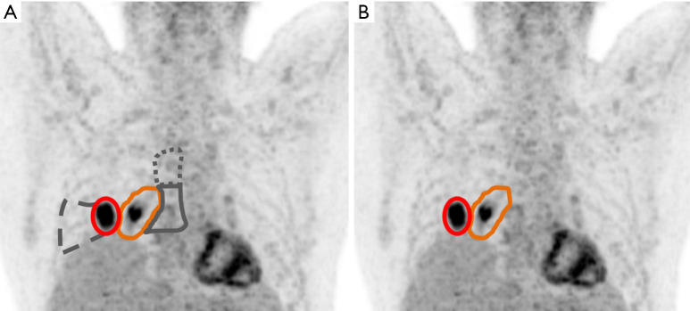
Radiotherapy (RT) target volume concepts for locally advanced lung cancer have been under discussion for years. Although they may be as important as treatment doses, many aspects of them are still based on conventions, which, due to the paucity of prospective data, rely on long-term practice or on clinical knowledge and experience (e.g., on patterns of spread or recurrence). However, in recent years, large improvements have been made in medical imaging and molecular imaging methods have been implemented, which are of great interest in RT. For lung cancer, in recent years, 18F-fluoro-desoxy-glucose (FDG)-positron-emission tomography (PET)/computed tomography (CT) has shown a superior diagnostic accuracy as compare to conventional imaging and has become an indispensable standard tool for diagnostic workup, staging and response assessment. This offers the chance to optimize target volume concepts in relation to modern imaging. While actual recommendations as the EORTC or ESTRO-ACROP guidelines already include imaging standards, the recently published PET-Plan trial prospectively investigated conventional versus imaging guided target volumes in relation to patient outcome. The results of this trial may help to further refine standards. The current review gives a practical overview on procedures for pre-treatment imaging and target volume delineation in locally advanced non-small cell lung cancer (NSCLC) in synopsis of the procedures established by the PET-Plan trial with the actual EORTC and ACROP guidelines.
局部晚期肺癌的放射治疗(RT)靶区概念多年来一直处于讨论之中。尽管它们可能与治疗剂量同样重要,但其许多方面仍基于惯例,由于前瞻性数据匮乏,这些惯例依赖长期实践或临床知识与经验(例如,基于扩散或复发模式)。然而,近年来,医学成像取得了巨大进步,分子成像方法也已得到应用,这在放射治疗中具有重要意义。对于肺癌,近年来,18F-氟脱氧葡萄糖(FDG)正电子发射断层扫描(PET)/计算机断层扫描(CT)与传统成像相比显示出更高的诊断准确性,并已成为诊断检查、分期和疗效评估不可或缺的标准工具。这为根据现代成像优化靶区概念提供了机会。虽然欧洲癌症研究与治疗组织(EORTC)或欧洲放射肿瘤学会-放射肿瘤学进展与合作组织(ESTRO-ACROP)指南等实际建议已经纳入了成像标准,但最近发表的PET-Plan试验前瞻性地研究了传统靶区与成像引导靶区对患者预后的影响。该试验结果可能有助于进一步完善标准。本综述结合PET-Plan试验以及实际的EORTC和ACROP指南所确立的程序,对局部晚期非小细胞肺癌(NSCLC)的治疗前成像和靶区勾画程序进行了实际概述。