Department of Neurology, Xuanwu Hospital, Capital Medical University, Beijing, China.
Advanced Center of Stroke, Beijing Institute for Brain Disorders, Beijing, China.
J Int Med Res. 2021 May;49(5):3000605211017001. doi: 10.1177/03000605211017001.
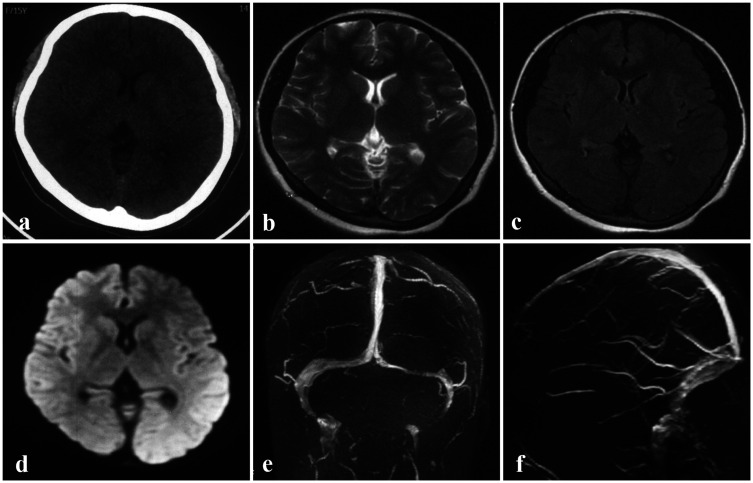
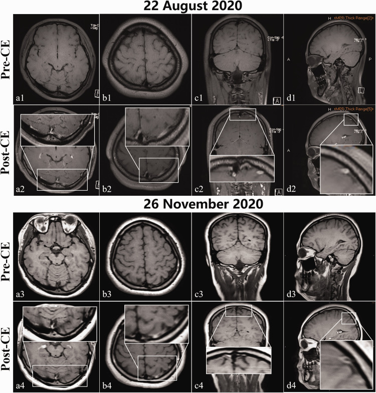
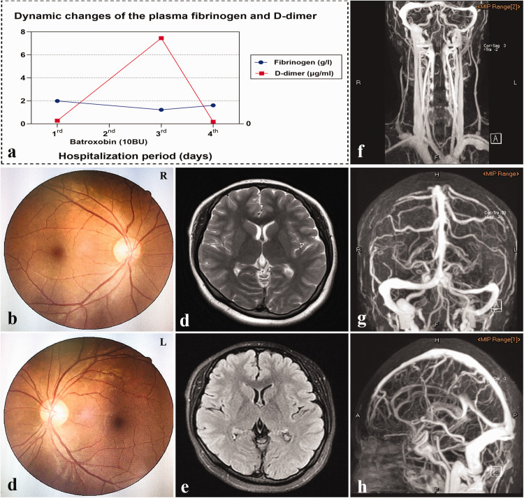
Cerebral venous thrombosis (CVT) is easily missed or misdiagnosed in clinical settings because of its high variability in terms of symptoms and radiological findings. Herein, we aimed to explore a promising modality for confirming presumed CVT in the hope to uncover its superior diagnostic performance to conventional imaging modalities. The patient complained of intolerable pain in her forehead and left eye. Her lumbar puncture opening pressure was 140 mmHO, and her cerebrospinal fluid composition was normal. No marked abnormalities were observed in routine brain images, including non-contrast computed tomography, magnetic resonance imaging, and contrast-enhanced magnetic resonance venography. However, chronic mural thrombi in the lumen of the left cortical veins, transverse/sigmoid sinus, and superior sagittal sinus were identified in magnetic resonance black-blood thrombus imaging (MRBTI) maps.
MRBTI can be used to directly and non-invasively visualize thrombi, and may thus be a promising tool over alternative routine techniques for confirming the diagnosis of CVT.
由于症状和影像学表现的高度多变性,脑静脉血栓形成(CVT)在临床环境中容易被忽视或误诊。在此,我们旨在探索一种有前途的方法来确认疑似 CVT,希望能发现其优于传统影像学方法的诊断性能。患者主诉前额和左眼剧痛。她的腰椎穿刺颅内压为 140mmHO,脑脊液成分正常。常规脑影像,包括非对比 CT、磁共振成像和对比增强磁共振静脉造影,均未见明显异常。然而,磁共振黑血血栓成像(MRBTI)图谱显示左侧皮质静脉、横窦/乙状窦和上矢状窦管腔内存在慢性壁血栓。
MRBTI 可用于直接、无创地显示血栓,因此可能是一种很有前途的工具,可以替代常规技术来确认 CVT 的诊断。