Goel Manvi, Mangel Stuart C
Department of Neuroscience, Ohio State University College of Medicine, Columbus, OH, United States.
Front Cell Neurosci. 2021 May 5;15:647541. doi: 10.3389/fncel.2021.647541. eCollection 2021.
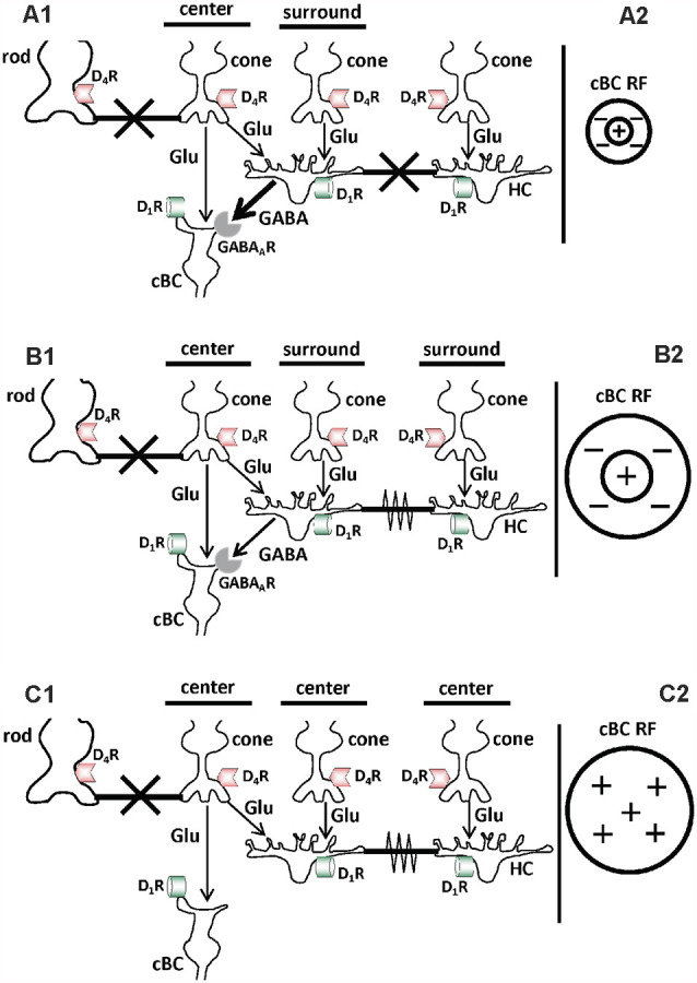
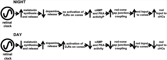
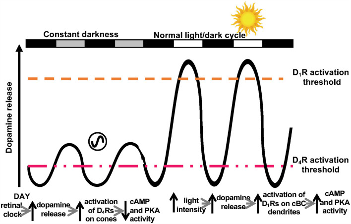
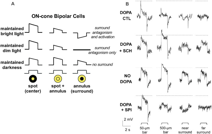
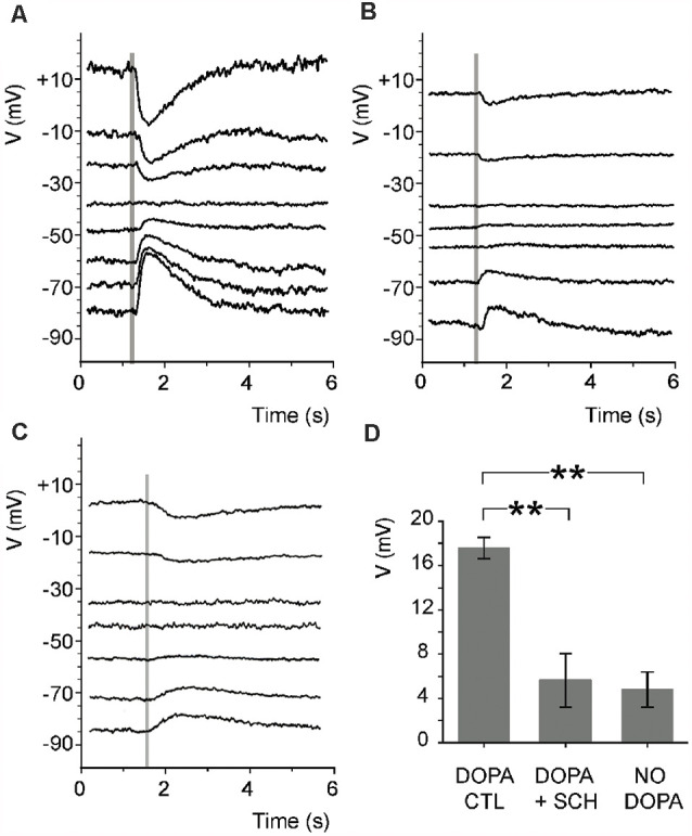
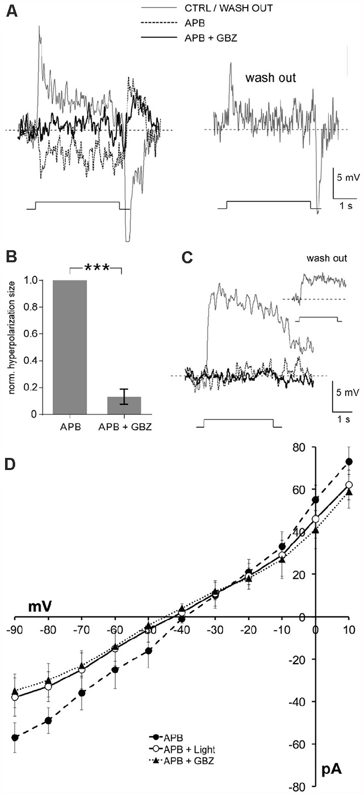
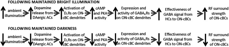
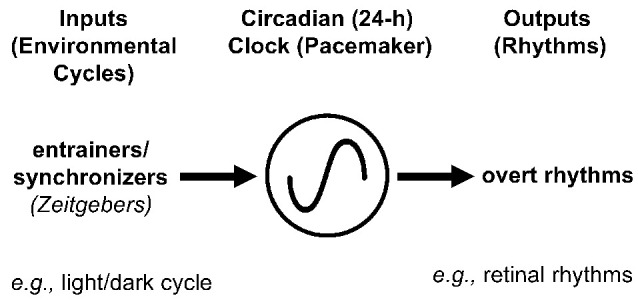
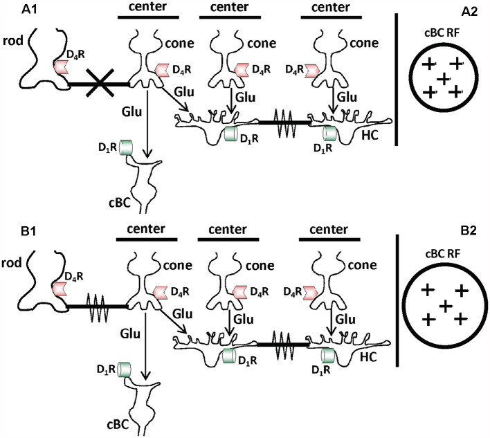
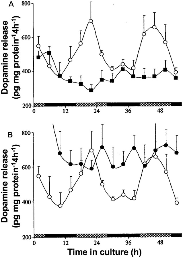
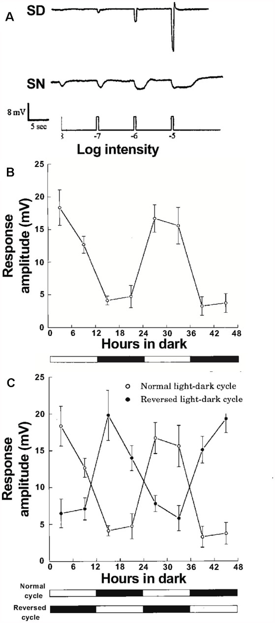
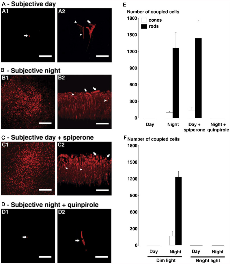
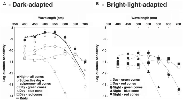
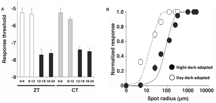
The vertebrate retina, like most other brain regions, undergoes relatively slow alterations in neural signaling in response to gradual changes in physiological conditions (e.g., activity changes to rest), or in response to gradual changes in environmental conditions (e.g., day changes into night). As occurs elsewhere in the brain, the modulatory processes that mediate slow adaptation in the retina are driven by extrinsic signals (e.g., changes in ambient light level) and/or by intrinsic signals such as those of the circadian (24-h) clock in the retina. This review article describes and discusses the extrinsic and intrinsic modulatory processes that enable neural circuits in the retina to optimize their visual performance throughout day and night as the ambient light level changes by ~10 billion-fold. In the first synaptic layer of the retina, cone photoreceptor cells form gap junctions with rods and signal cone-bipolar and horizontal cells (HCs). Distinct extrinsic and intrinsic modulatory processes in this synaptic layer are mediated by long-range feedback of the neuromodulator dopamine. Dopamine is released by dopaminergic cells, interneurons whose cell bodies are located in the second synaptic layer of the retina. Distinct actions of dopamine modulate chemical and electrical synapses in day and night. The retinal circadian clock increases dopamine release in the day compared to night, activating high-affinity dopamine D receptors on cones. This clock effect controls electrical synapses between rods and cones so that rod-cone electrical coupling is minimal in the day and robust at night. The increase in rod-cone coupling at night improves the signal-to-noise ratio and the reliability of very dim multi-photon light responses, thereby enhancing detection of large dim objects on moonless nights.Conversely, maintained (30 min) bright illumination in the day compared to maintained darkness releases sufficient dopamine to activate low-affinity dopamine D receptors on cone-bipolar cell dendrites. This non-circadian light/dark adaptive process regulates the function of GABA receptors on ON-cone-bipolar cell dendrites so that the receptive field (RF) surround of the cells is strong following maintained bright illumination but minimal following maintained darkness. The increase in surround strength in the day following maintained bright illumination enhances the detection of edges and fine spatial details.
脊椎动物的视网膜与大多数其他脑区一样,会随着生理状况的逐渐变化(例如,从活动状态转变为休息状态)或环境条件的逐渐变化(例如,从白天转变为夜晚),在神经信号传递方面发生相对缓慢的改变。正如在大脑其他部位所发生的情况一样,介导视网膜缓慢适应的调节过程由外部信号(例如,环境光水平的变化)和/或内部信号驱动,如视网膜中昼夜节律(24小时)时钟发出的信号。这篇综述文章描述并讨论了外部和内部调节过程,这些过程使视网膜中的神经回路能够随着环境光水平变化约100亿倍,在白天和黑夜全天优化其视觉性能。在视网膜的第一个突触层中,视锥光感受器细胞与视杆细胞形成缝隙连接,并向视锥双极细胞和水平细胞(HCs)发送信号。该突触层中不同的外部和内部调节过程由神经调质多巴胺的长程反馈介导。多巴胺由多巴胺能细胞释放,这些中间神经元的细胞体位于视网膜的第二个突触层。多巴胺的不同作用在白天和黑夜调节化学突触和电突触。与夜晚相比,视网膜昼夜节律时钟在白天增加多巴胺释放,激活视锥细胞上的高亲和力多巴胺D受体。这种时钟效应控制视杆细胞和视锥细胞之间的电突触,使得视杆 - 视锥电耦合在白天最小,而在夜晚很强。夜晚视杆 - 视锥耦合的增加提高了信噪比和非常微弱的多光子光反应的可靠性,从而增强了在无月夜对大型暗物体的检测。相反,与持续黑暗相比,白天持续(30分钟)明亮光照会释放足够的多巴胺,以激活视锥双极细胞树突上的低亲和力多巴胺D受体。这种非昼夜的光/暗适应过程调节视锥双极细胞树突上GABA受体的功能,使得在持续明亮光照后,这些细胞的感受野(RF)周围很强,但在持续黑暗后最小。在白天持续明亮光照后,周围强度的增加增强了对边缘和精细空间细节的检测。