Zhang Lu, Fang Yu, Zhao Xinyu, Zheng Yake, Ma Yunqing, Li Shuang, Huang Zhi, Li Lihao
Department of Neurology, the First Affiliated Hospital of Zhengzhou University, Zhengzhou 450052, P.R. China.
ICU, The First Affiliated Hospital of Zhengzhou University, Zhengzhou 450052, P.R. China.
Mol Ther Nucleic Acids. 2021 Feb 15;24:822-831. doi: 10.1016/j.omtn.2021.02.010. eCollection 2021 Jun 4.
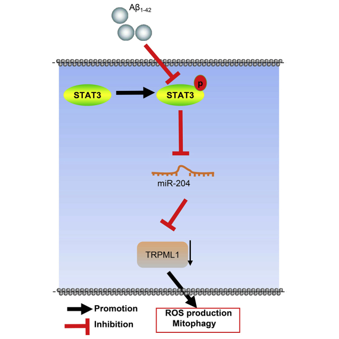
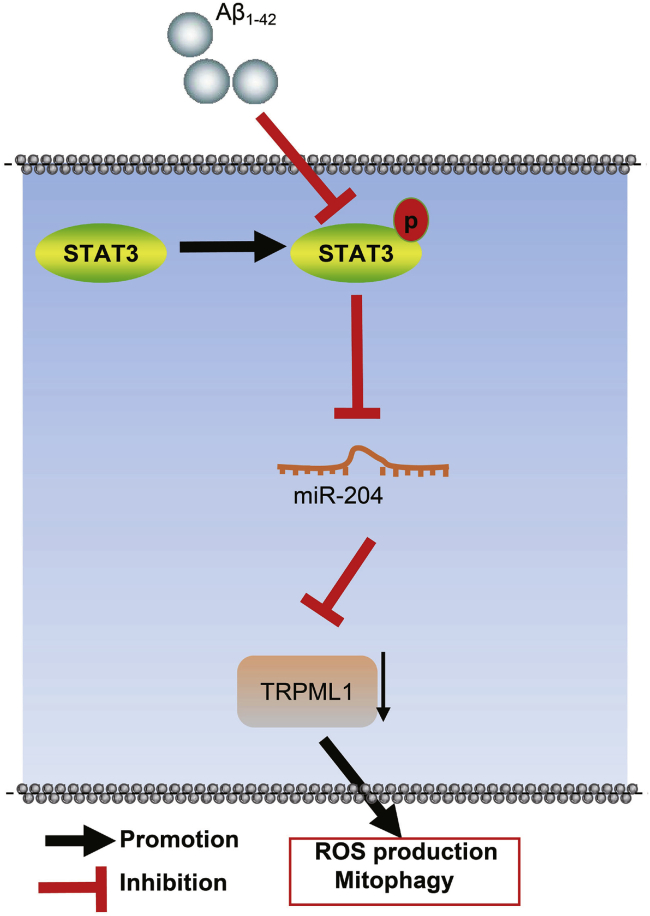
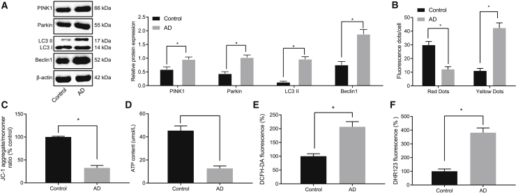
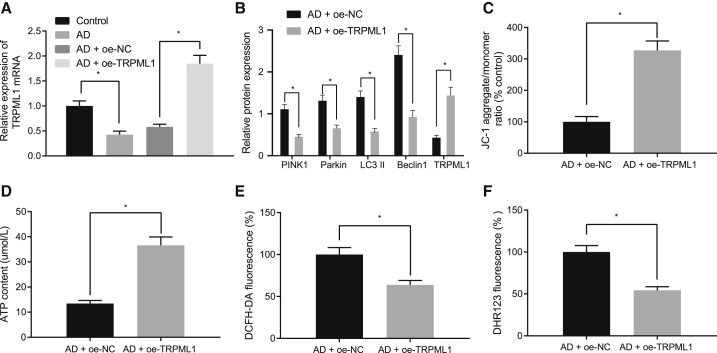
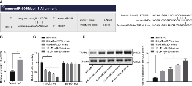
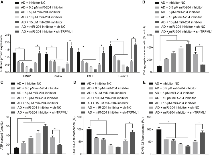
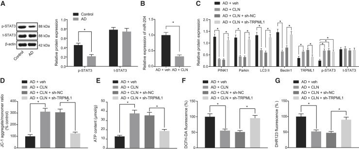
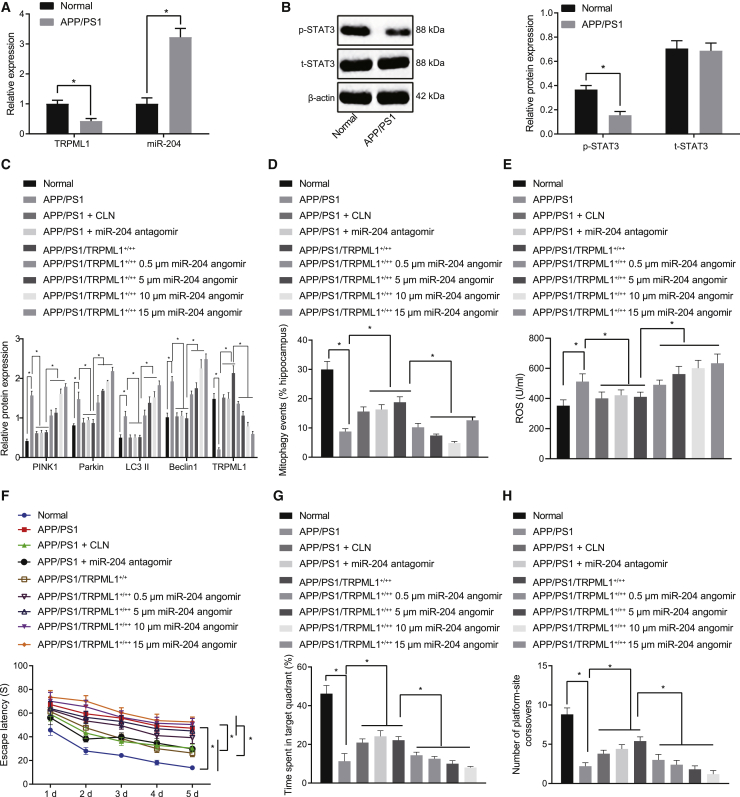
Mitochondrial dysfunction is an early feature of Alzheimer's disease (AD), whereby accumulation of damaged mitochondria in conjunction with impaired mitophagy contributes to neurodegeneration. Various non-transcribed microRNAs (miRNAs) are involved in this process. In the present study, we aimed to decipher the participation of miR-204 in a murine AD model. Primary hippocampal neurons were isolated from mice and treated with β-amyloid 1-42 (Aβ1-42) to establish a cell model of AD. Dichloro-dihydro-fluorescein diacetate and dihydrorhodamine 123 staining assays were performed to measure total reactive oxygen species (ROS) and mitochondrial ROS production in neurons, and MitoSOX staining was done to analyze mitochondrial ROS production in hippocampus. Furthermore, mitochondrial autophagy was observed in hippocampus from amyloid precursor protein/pesenilin-1 AD modeled mice, and their cognitive function was assessed by Morris water maze. Mitochondrial damage, ROS production, and mitochondrial autophagy were observed in AD cell model induced by Aβ1-42. In AD, signal transducer and activator of transcription 3 (STAT3) and transient receptor potential mucolipin-1 (TRPML1) expression was downregulated, although miR-204 expression was upregulated. TRPML1 overexpression, downregulation of miR-204, or STAT3 pathway activation reduced the Aβ1-42-induced mitochondrial damage, along with ROS production and mitochondrial autophagy and . Silencing of miR-204 could upregulate TRPML1 expression, thus suppressing ROS production and mitochondrial autophagy in AD through STAT3 pathway.
线粒体功能障碍是阿尔茨海默病(AD)的早期特征,受损线粒体的积累与线粒体自噬受损共同导致神经退行性变。多种非编码微小RNA(miRNA)参与了这一过程。在本研究中,我们旨在阐明miR-204在小鼠AD模型中的作用。从小鼠中分离出原代海马神经元,并用β-淀粉样蛋白1-42(Aβ1-42)处理以建立AD细胞模型。进行二氯二氢荧光素二乙酸酯和二氢罗丹明123染色试验以测量神经元中的总活性氧(ROS)和线粒体ROS生成,并进行MitoSOX染色以分析海马中的线粒体ROS生成。此外,在淀粉样前体蛋白/早老素-1 AD模型小鼠的海马中观察线粒体自噬,并通过莫里斯水迷宫评估其认知功能。在Aβ1-42诱导的AD细胞模型中观察到线粒体损伤、ROS生成和线粒体自噬。在AD中,信号转导和转录激活因子3(STAT3)和瞬时受体电位黏蛋白1(TRPML1)的表达下调,尽管miR-204表达上调。TRPML1过表达、miR-204下调或STAT3途径激活可减少Aβ1-42诱导的线粒体损伤以及ROS生成和线粒体自噬。miR-204沉默可上调TRPML1表达,从而通过STAT3途径抑制AD中的ROS生成和线粒体自噬。