Normandie Univ, UNICAEN, CEA, CNRS, ISTCT/CERVOxy Group, GIP CYCERON, 14000, Caen, France.
Medical Physics Department, CLCC François Baclesse, 14000, Caen, France.
Sci Rep. 2021 May 27;11(1):11239. doi: 10.1038/s41598-021-90662-0.
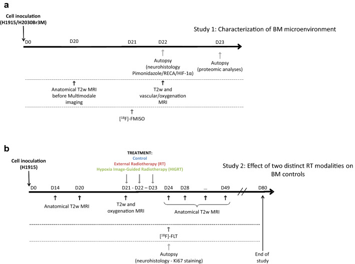
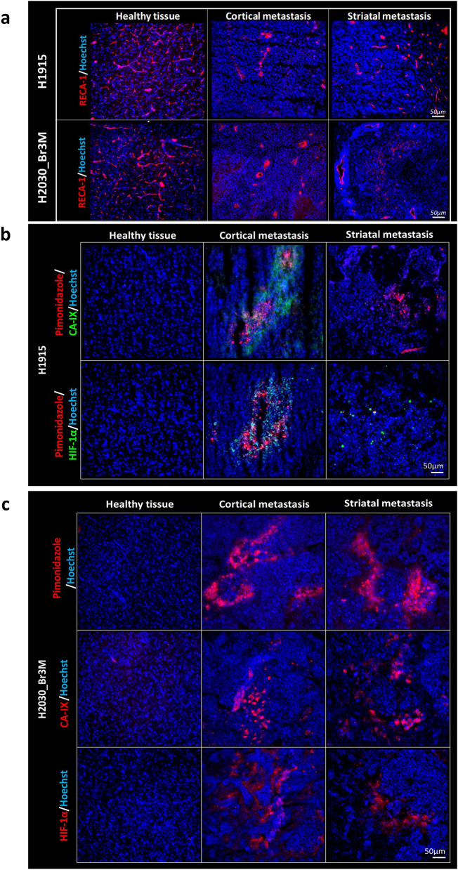
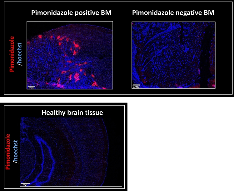
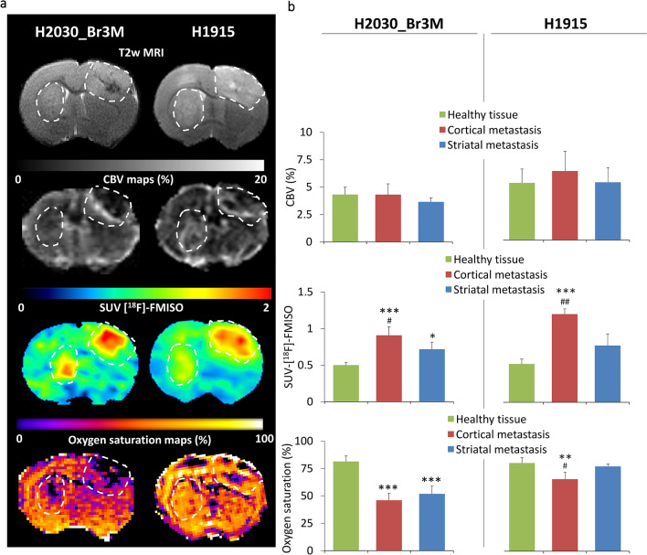
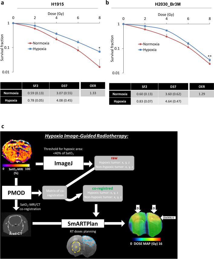
Lung cancer patients frequently develop brain metastases (BM). Despite aggressive treatment including neurosurgery and external-radiotherapy, overall survival remains poor. There is a pressing need to further characterize factors in the microenvironment of BM that may confer resistance to radiotherapy (RT), such as hypoxia. Here, hypoxia was first evaluated in 28 biopsies from patients with non‑small cell lung cancer (NSCLC) BM, using CA-IX immunostaining. Hypoxia characterization (pimonidazole, CA-IX and HIF-1α) was also performed in different preclinical NSCLC BM models induced either by intracerebral injection of tumor cells (H2030-Br3M, H1915) into the cortex and striatum, or intracardial injection of tumor cells (H2030-Br3M). Additionally, [F]-FMISO-PET and oxygen-saturation-mapping-MRI (SatO2-MRI) were carried out in the intracerebral BM models to further characterize tumor hypoxia and evaluate the potential of Hypoxia-image-guided-RT (HIGRT). The effect of RT on proliferation of BM ([F]-FLT-PET), tumor volume and overall survival was determined. We showed that hypoxia is a major yet heterogeneous feature of BM from lung cancer both preclinically and clinically. HIGRT, based on hypoxia heterogeneity observed between cortical and striatal metastases in the intracerebrally induced models, showed significant potential for tumor control and animal survival. These results collectively highlight hypoxia as a hallmark of BM from lung cancer and the value of HIGRT in better controlling tumor growth.
肺癌患者常发生脑转移(BM)。尽管包括神经外科手术和外放射治疗在内的积极治疗,总体生存率仍然较差。进一步明确 BM 微环境中可能对放射治疗(RT)产生抵抗的因素(如缺氧)具有迫切的需要。本研究首次通过 CA-IX 免疫染色评估了 28 例非小细胞肺癌(NSCLC)BM 患者的活检标本中的缺氧情况。还在通过肿瘤细胞脑内注射(H2030-Br3M、H1915)到皮质和纹状体或心脏内注射肿瘤细胞(H2030-Br3M)诱导的不同 NSCLC BM 临床前模型中进行了缺氧特征(pimonidazole、CA-IX 和 HIF-1α)的描述。此外,在脑内 BM 模型中还进行了 [F]-FMISO-PET 和氧饱和度映射 MRI(SatO2-MRI),以进一步描述肿瘤缺氧情况并评估缺氧成像引导放疗(HIGRT)的潜力。本研究还检测了 RT 对 BM 增殖([F]-FLT-PET)、肿瘤体积和总体生存的影响。我们发现,缺氧是肺癌临床前和临床 BM 的一个主要的异质性特征。基于脑内诱导模型中皮质和纹状体转移之间观察到的缺氧异质性的 HIGRT,显示出对肿瘤控制和动物生存有显著的潜力。这些结果共同强调了缺氧是肺癌 BM 的一个标志,以及 HIGRT 在更好地控制肿瘤生长方面的价值。