• 文献检索
  • 文档翻译
  • 深度研究
  • 学术资讯
  • Suppr Zotero 插件Zotero 插件
  • 邀请有礼
  • 套餐&价格
  • 历史记录
应用&插件
Suppr Zotero 插件Zotero 插件浏览器插件Mac 客户端Windows 客户端微信小程序
定价
高级版会员购买积分包购买API积分包
服务
文献检索文档翻译深度研究API 文档MCP 服务
关于我们
关于 Suppr公司介绍联系我们用户协议隐私条款
关注我们

Suppr 超能文献

核心技术专利:CN118964589B侵权必究
粤ICP备2023148730 号-1Suppr @ 2026

文献检索

告别复杂PubMed语法,用中文像聊天一样搜索,搜遍4000万医学文献。AI智能推荐,让科研检索更轻松。

立即免费搜索

文件翻译

保留排版,准确专业,支持PDF/Word/PPT等文件格式,支持 12+语言互译。

免费翻译文档

深度研究

AI帮你快速写综述,25分钟生成高质量综述,智能提取关键信息,辅助科研写作。

立即免费体验

脾脏感染的横断面成像表现:能否进行鉴别诊断?

Cross-sectional imaging findings of splenic infections: is differential diagnosis possible?

机构信息

Department of Radiology, Hacettepe University School of Medicine, Ankara, 06100, Turkey.

Department of Radiology, Gulhane Training and Research Hospital, Ankara, 06010, Turkey.

出版信息

Abdom Radiol (NY). 2021 Oct;46(10):4828-4852. doi: 10.1007/s00261-021-03130-8. Epub 2021 May 28.

DOI:10.1007/s00261-021-03130-8
PMID:34047800
原文链接:https://pmc.ncbi.nlm.nih.gov/articles/PMC8160561/
Abstract

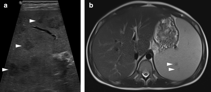
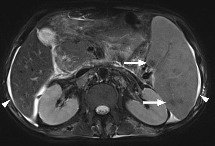
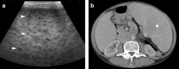
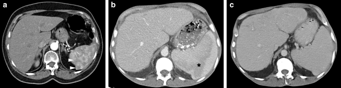
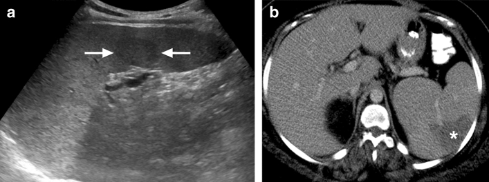
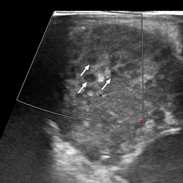
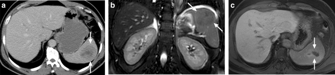
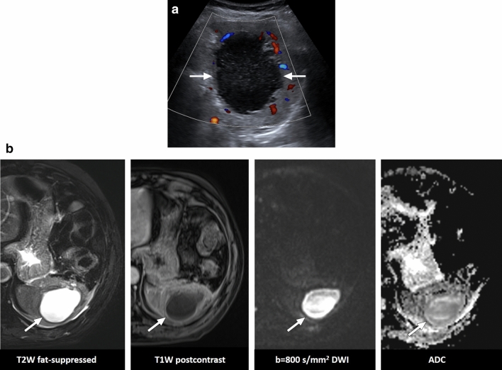
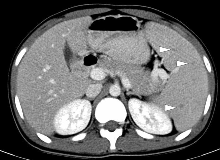
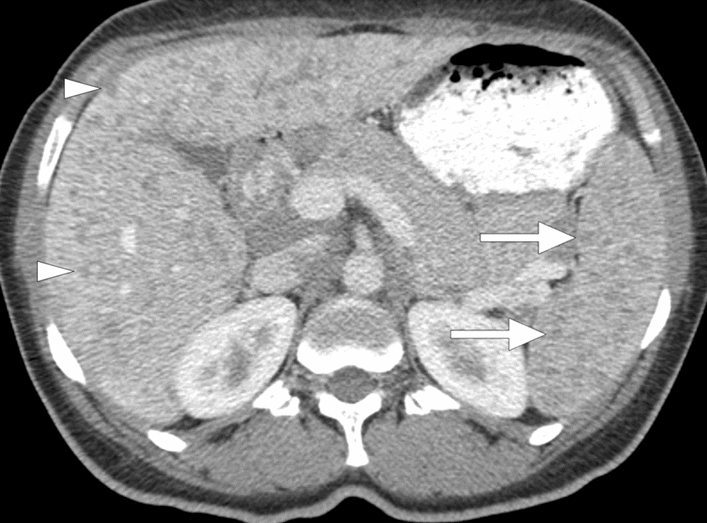
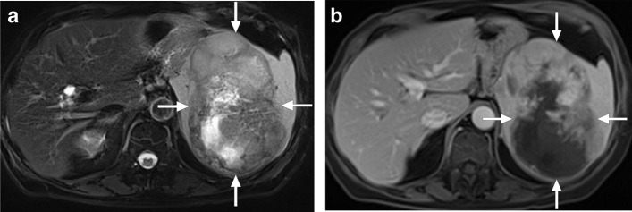
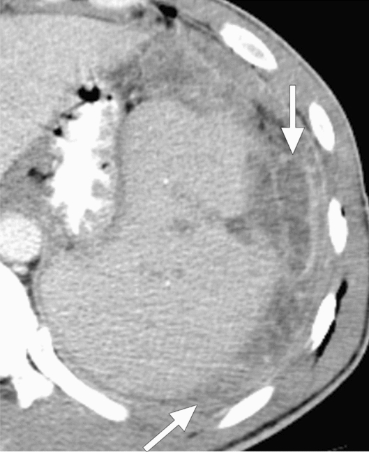
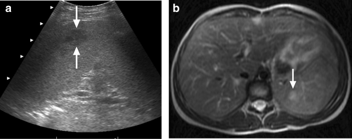
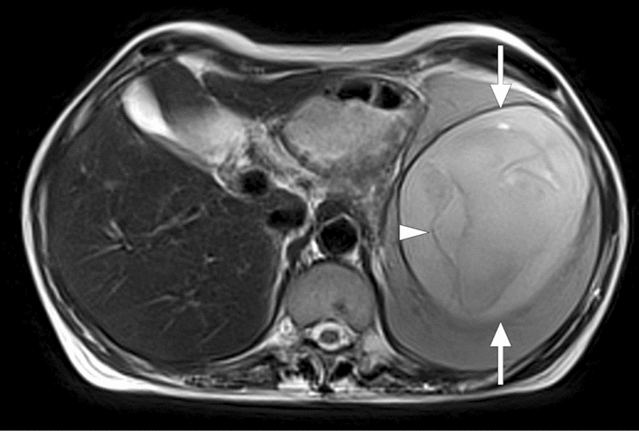
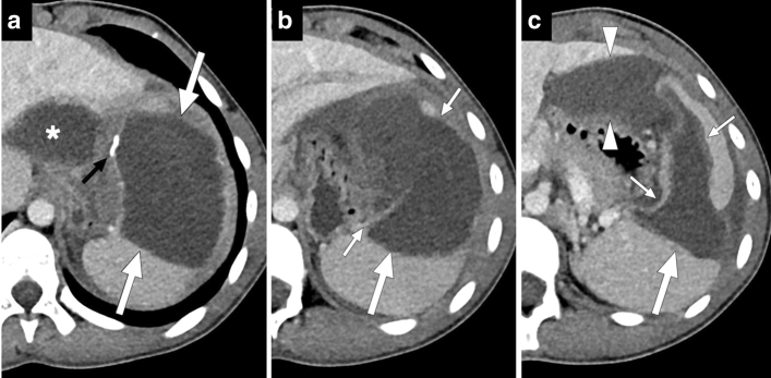
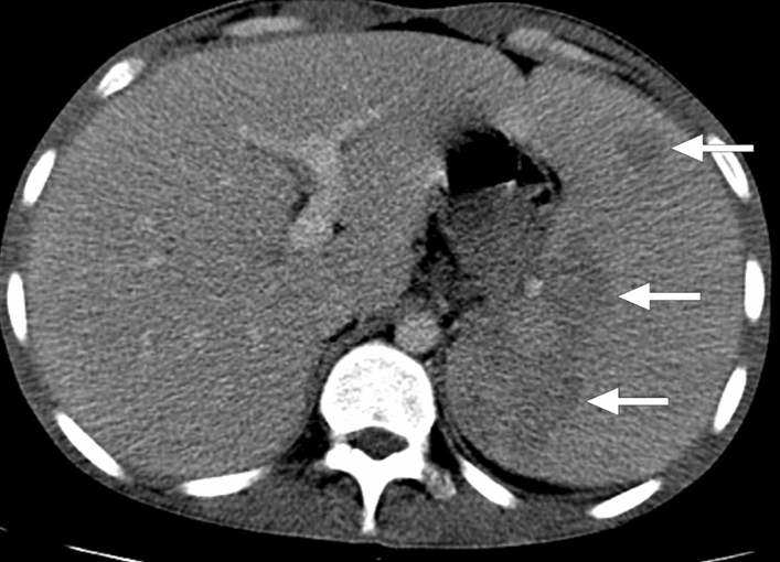
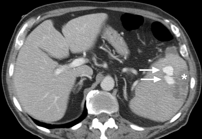
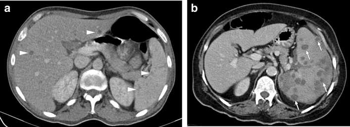
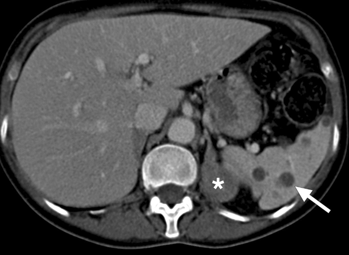
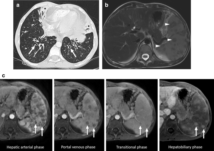
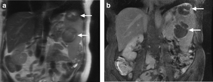
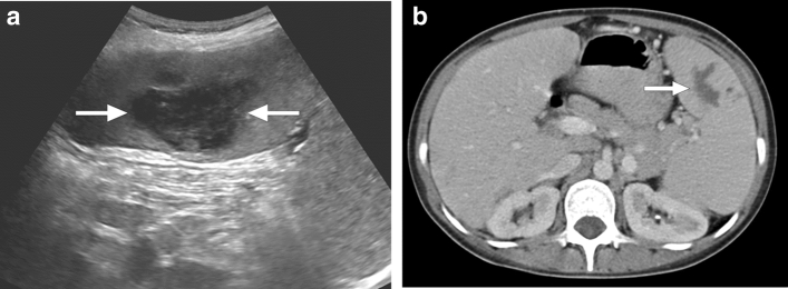
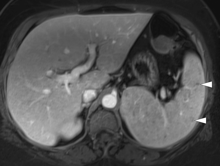
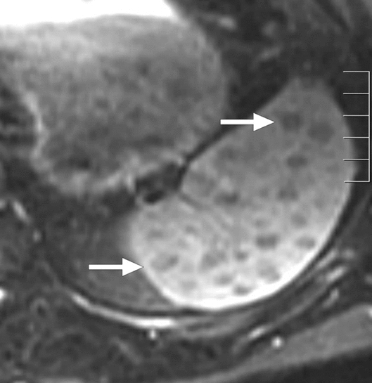
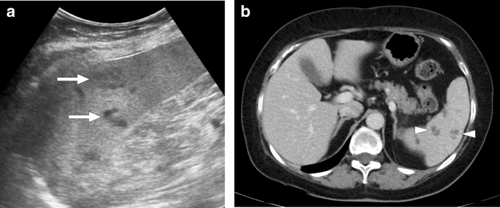
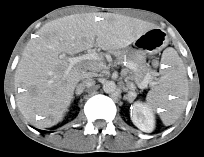
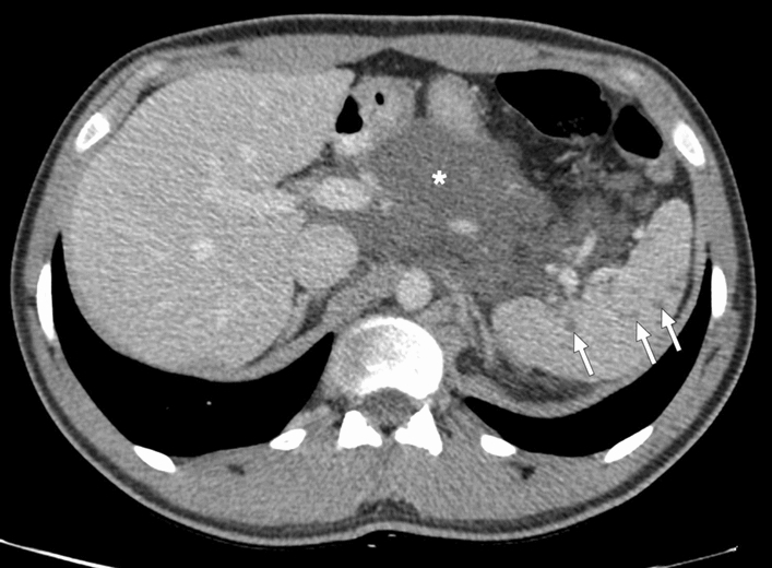
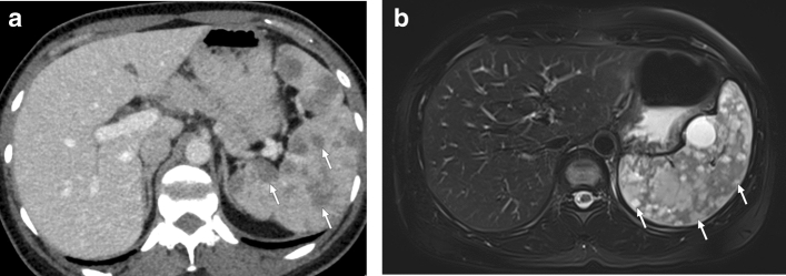
The spleen plays an important role in the immunological homeostasis of the body. Several neoplastic and non-neoplastic diseases may affect this organ, and imaging is of fundamental importance for diagnosis. Infectious diseases of the spleen can be encountered in daily radiology practice, and differential diagnosis may sometimes be challenging. Infectious involvement of the spleen can be primary or secondary to a different source outside the spleen. Despite the fact that different infectious diseases may cause similar imaging findings, we believe that differential diagnosis between different causes may also be possible in certain patients with imaging. Early diagnosis may potentially enhance patients' treatment and outcome. In this review, we aimed to increase imaging specialists' awareness of splenic infections by describing the multimodality imaging features of common and atypical infections of the spleen with their differential diagnoses.

摘要

脾脏在人体的免疫稳态中发挥着重要作用。几种肿瘤性和非肿瘤性疾病可能会影响到这个器官,而影像学对于诊断具有重要意义。脾脏的感染性疾病在日常放射学实践中较为常见,鉴别诊断有时可能具有挑战性。脾脏的感染性累及可以是原发性的,也可以是脾脏以外的其他部位的感染性疾病的继发性累及。尽管不同的感染性疾病可能会引起相似的影像学表现,但我们认为,在某些具有影像学表现的患者中,不同病因之间的鉴别诊断也是可能的。早期诊断可能会潜在地改善患者的治疗和预后。在这篇综述中,我们旨在通过描述脾脏常见和非典型感染的多模态影像学特征及其鉴别诊断,提高影像专家对脾脏感染的认识。

https://cdn.ncbi.nlm.nih.gov/pmc/blobs/0f0f/8160561/75c56e100a6f/261_2021_3130_Fig29_HTML.jpg
https://cdn.ncbi.nlm.nih.gov/pmc/blobs/0f0f/8160561/739b97b32186/261_2021_3130_Fig1_HTML.jpg
https://cdn.ncbi.nlm.nih.gov/pmc/blobs/0f0f/8160561/01df9ceda2d1/261_2021_3130_Fig2_HTML.jpg
https://cdn.ncbi.nlm.nih.gov/pmc/blobs/0f0f/8160561/3a97c9ac59b4/261_2021_3130_Fig3_HTML.jpg
https://cdn.ncbi.nlm.nih.gov/pmc/blobs/0f0f/8160561/369b03a70e88/261_2021_3130_Fig4_HTML.jpg
https://cdn.ncbi.nlm.nih.gov/pmc/blobs/0f0f/8160561/3322787c80ab/261_2021_3130_Fig5_HTML.jpg
https://cdn.ncbi.nlm.nih.gov/pmc/blobs/0f0f/8160561/d85327c6c277/261_2021_3130_Fig6_HTML.jpg
https://cdn.ncbi.nlm.nih.gov/pmc/blobs/0f0f/8160561/70ba992c00f8/261_2021_3130_Fig7_HTML.jpg
https://cdn.ncbi.nlm.nih.gov/pmc/blobs/0f0f/8160561/33a62e352265/261_2021_3130_Fig8_HTML.jpg
https://cdn.ncbi.nlm.nih.gov/pmc/blobs/0f0f/8160561/1ea3f051b979/261_2021_3130_Fig9_HTML.jpg
https://cdn.ncbi.nlm.nih.gov/pmc/blobs/0f0f/8160561/581a38afbf5a/261_2021_3130_Fig10_HTML.jpg
https://cdn.ncbi.nlm.nih.gov/pmc/blobs/0f0f/8160561/ccf0a0b4866f/261_2021_3130_Fig11_HTML.jpg
https://cdn.ncbi.nlm.nih.gov/pmc/blobs/0f0f/8160561/12a1c7330d00/261_2021_3130_Fig12_HTML.jpg
https://cdn.ncbi.nlm.nih.gov/pmc/blobs/0f0f/8160561/c7766193178c/261_2021_3130_Fig13_HTML.jpg
https://cdn.ncbi.nlm.nih.gov/pmc/blobs/0f0f/8160561/9c86d1f7190a/261_2021_3130_Fig14_HTML.jpg
https://cdn.ncbi.nlm.nih.gov/pmc/blobs/0f0f/8160561/bf14642cb9a7/261_2021_3130_Fig15_HTML.jpg
https://cdn.ncbi.nlm.nih.gov/pmc/blobs/0f0f/8160561/09e03fb7cbe3/261_2021_3130_Fig16_HTML.jpg
https://cdn.ncbi.nlm.nih.gov/pmc/blobs/0f0f/8160561/e8b74a25278e/261_2021_3130_Fig17_HTML.jpg
https://cdn.ncbi.nlm.nih.gov/pmc/blobs/0f0f/8160561/242c7707b746/261_2021_3130_Fig18_HTML.jpg
https://cdn.ncbi.nlm.nih.gov/pmc/blobs/0f0f/8160561/ea4642c8226d/261_2021_3130_Fig19_HTML.jpg
https://cdn.ncbi.nlm.nih.gov/pmc/blobs/0f0f/8160561/155f1ba891c7/261_2021_3130_Fig20_HTML.jpg
https://cdn.ncbi.nlm.nih.gov/pmc/blobs/0f0f/8160561/d31a7cdc3ab0/261_2021_3130_Fig21_HTML.jpg
https://cdn.ncbi.nlm.nih.gov/pmc/blobs/0f0f/8160561/67757e94bee3/261_2021_3130_Fig22_HTML.jpg
https://cdn.ncbi.nlm.nih.gov/pmc/blobs/0f0f/8160561/0c131bb4f6e9/261_2021_3130_Fig23_HTML.jpg
https://cdn.ncbi.nlm.nih.gov/pmc/blobs/0f0f/8160561/fb2f01f5b257/261_2021_3130_Fig24_HTML.jpg
https://cdn.ncbi.nlm.nih.gov/pmc/blobs/0f0f/8160561/bf71fc6e984a/261_2021_3130_Fig25_HTML.jpg
https://cdn.ncbi.nlm.nih.gov/pmc/blobs/0f0f/8160561/55c92d078ee2/261_2021_3130_Fig26_HTML.jpg
https://cdn.ncbi.nlm.nih.gov/pmc/blobs/0f0f/8160561/1f7d7da5935a/261_2021_3130_Fig27_HTML.jpg
https://cdn.ncbi.nlm.nih.gov/pmc/blobs/0f0f/8160561/75c56e100a6f/261_2021_3130_Fig29_HTML.jpg

相似文献

1
Cross-sectional imaging findings of splenic infections: is differential diagnosis possible?脾脏感染的横断面成像表现:能否进行鉴别诊断?
Abdom Radiol (NY). 2021 Oct;46(10):4828-4852. doi: 10.1007/s00261-021-03130-8. Epub 2021 May 28.
2
[Diagnostic imaging of splenic disease].[脾脏疾病的诊断成像]
Radiologe. 2006 Mar;46(3):229-43; quiz 244. doi: 10.1007/s00117-005-1333-8.
3
Nonneoplastic, benign, and malignant splenic diseases: cross-sectional imaging findings and rare disease entities.非肿瘤性、良性和恶性脾疾病:影像学表现及罕见病种。
AJR Am J Roentgenol. 2014 Aug;203(2):315-22. doi: 10.2214/AJR.13.11777.
4
Splenic sarcoidosis mimicking neoplastic disease.酷似肿瘤性疾病的脾结节病
J Clin Ultrasound. 2014 Jan;42(1):38-41. doi: 10.1002/jcu.22031. Epub 2013 Jan 4.
5
Hot spleen: hypervascular lesions of the spleen.热脾:脾脏的高血运性病变。
Abdom Imaging. 2015 Oct;40(7):2796-813. doi: 10.1007/s00261-015-0523-8.
6
Improving diagnosis of atraumatic splenic lesions, part II: benign neoplasms/nonneoplastic mass-like lesions.提高非创伤性脾病变的诊断水平,第二部分:良性肿瘤/非肿瘤性肿块样病变
Clin Imaging. 2016 Jul-Aug;40(4):691-704. doi: 10.1016/j.clinimag.2016.02.002. Epub 2016 Feb 6.
7
Multiple lesions of the spleen: differential diagnosis of cystic and solid lesions.脾脏的多发性病变:囊性和实性病变的鉴别诊断
Semin Ultrasound CT MR. 2006 Oct;27(5):389-403. doi: 10.1053/j.sult.2006.06.004.
8
MDCT Findings of Splenic Pathology.脾脏病变的 MDCT 表现。
Curr Probl Diagn Radiol. 2022 Mar-Apr;51(2):262-269. doi: 10.1067/j.cpradiol.2020.12.004. Epub 2021 Jan 9.
9
Sonography of the spleen.脾脏超声检查。
J Ultrasound Med. 2011 Sep;30(9):1281-93. doi: 10.7863/jum.2011.30.9.1281.
10
Splenic hydatidosis: a rare differential diagnosis in a cystic lesion of the spleen.脾包虫病:脾脏囊性病变中一种罕见的鉴别诊断。
Rev Gastroenterol Mex. 2007 Apr-Jun;72(2):122-5.

引用本文的文献

1
Reactivation of Latent Tuberculosis Following COVID-19 and Epstein-Barr Virus Coinfection: A Case Report.新型冠状病毒肺炎与爱泼斯坦-巴尔病毒合并感染后潜伏性结核病的再激活:一例报告
Pathogens. 2025 May 16;14(5):488. doi: 10.3390/pathogens14050488.
2
Multidisciplinary Management of Single Organism Emphysematous Splenitis Without Splenectomy: A Case Report.非脾切除术治疗单微生物气肿性脾炎的多学科管理:一例报告
Cureus. 2025 Mar 11;17(3):e80389. doi: 10.7759/cureus.80389. eCollection 2025 Mar.
3
Multimodality Imaging Features of Various Splenic Lesions: Clinical and Histopathologic Correlation.

本文引用的文献

1
Splenic infarction and infectious diseases in Korea.韩国的脾梗死与传染性疾病。
BMC Infect Dis. 2020 Dec 2;20(1):915. doi: 10.1186/s12879-020-05645-9.
2
Disseminated Nocardiosis with Lung, Liver and Spleen Abscesses in Sweet Syndrome: A Case Report.Sweet综合征合并肺部、肝脏和脾脏脓肿的播散性诺卡菌病:一例报告
J Acute Med. 2017 Dec 1;7(4):167-170. doi: 10.6705/j.jacme.2017.0704.005.
3
Case Report: Sudden Splenic Rupture in a -Infected Patient.病例报告:一名感染患者突发脾破裂。
各种脾脏病变的多模态成像特征:临床与组织病理学相关性
J Korean Soc Radiol. 2024 Nov;85(6):1099-1125. doi: 10.3348/jksr.2024.0004. Epub 2024 Nov 22.
4
[Infarction and splenic abscess as the cause of acute abdomen in a patient with myelomonocytic leukemia].[梗死和脾脓肿作为一名骨髓单核细胞白血病患者急性腹痛的病因]
Rev Med Inst Mex Seguro Soc. 2024 May 6;62(3):1-5. doi: 10.5281/zenodo.10999019.
5
Imaging and clinical manifestations of hematogenous dissemination in melioidosis.血源性播散性类鼻疽病的影像学表现及临床特征。
BMC Med Imaging. 2024 Nov 1;24(1):296. doi: 10.1186/s12880-024-01471-6.
6
Spleen anomalies and lesions in CT and MRI: essentials for radiologists and clinicians-a pictorial review.CT和MRI检查中的脾脏异常与病变:放射科医生和临床医生必备知识——图文综述
Abdom Radiol (NY). 2025 Feb;50(2):860-874. doi: 10.1007/s00261-024-04405-6. Epub 2024 Aug 28.
7
The Spectrum of Solitary Benign Splenic Lesions-Imaging Clues for a Noninvasive Diagnosis.孤立性脾脏良性病变的谱——无创诊断的影像学线索
Diagnostics (Basel). 2023 Jun 20;13(12):2120. doi: 10.3390/diagnostics13122120.
8
Splenic Abscess in the New Millennium: A Descriptive, Retrospective Case Series.新千年的脾脓肿:一项描述性回顾性病例系列研究。
Open Forum Infect Dis. 2022 Feb 17;9(4):ofac085. doi: 10.1093/ofid/ofac085. eCollection 2022 Apr.
Int J Gen Med. 2020 Sep 9;13:595-598. doi: 10.2147/IJGM.S267197. eCollection 2020.
4
Spectrum of Imaging Findings at Chest Radiography, US, CT, and MRI in Multisystem Inflammatory Syndrome in Children Associated with COVID-19.COVID-19 相关儿童多系统炎症综合征的胸部 X 线摄影、US、CT 和 MRI 的影像学表现谱。
Radiology. 2021 Jan;298(1):E1-E10. doi: 10.1148/radiol.2020202543. Epub 2020 Jun 25.
5
A Rare Case of Splenic in a HIV-Positive Patient.一名HIV阳性患者的罕见脾脏病例。 (原文“Splenic”后缺少具体病症等关键信息,翻译可能存在一定局限性)
Case Rep Gastrointest Med. 2020 Jun 10;2020:8509591. doi: 10.1155/2020/8509591. eCollection 2020.
6
Systemic Melioidosis With Ruptured Splenic Abscess.伴有脾脓肿破裂的全身性类鼻疽病
Cureus. 2020 May 4;12(5):e7956. doi: 10.7759/cureus.7956.
7
Calcified Splenic Lesions: Pattern Recognition Approach on CT With Pathologic Correlation.钙化性脾脏病变:CT 表现与病理对照的模式识别方法。
AJR Am J Roentgenol. 2020 May;214(5):1083-1091. doi: 10.2214/AJR.19.22246. Epub 2020 Mar 24.
8
Re-drawing the Maps for Endemic Mycoses.重新绘制地方病真菌图谱。
Mycopathologia. 2020 Oct;185(5):843-865. doi: 10.1007/s11046-020-00431-2. Epub 2020 Feb 10.
9
Splenic findings in patients with acute babesiosis.急性巴贝斯虫病患者的脾脏表现。
Abdom Radiol (NY). 2020 Mar;45(3):710-715. doi: 10.1007/s00261-019-02362-z.
10
Spontaneous Splenic Rupture Complicating Severe Infection: A Case Report and Literature Review.自发性脾破裂并发严重感染:一例报告及文献复习
Case Rep Infect Dis. 2019 Oct 24;2019:2781647. doi: 10.1155/2019/2781647. eCollection 2019.