Berlin Ultrahigh Field Facility (B.U.F.F.), Max Delbrück Center for Molecular Medicine in the Helmholtz Association (MDC), Robert-Rössle Strasse 10, 13125, Berlin, Germany.
DZHK (German Centre for Cardiovascular Research), Partner site Berlin, Berlin, Germany.
J Cardiovasc Magn Reson. 2021 May 31;23(1):63. doi: 10.1186/s12968-021-00754-z.
Hypertrophic cardiomyopathy (HCM) related myocardial vascular remodelling may lead to the reduction of myocardial blood supply and a subsequent progressive loss of cardiac function. This process has been difficult to observe and thus their connection remains unclear. Here we used non-invasive myocardial blood flow sensitive CMR to show an impairment of resting myocardial perfusion in a mouse model of naturally occurring HCM.
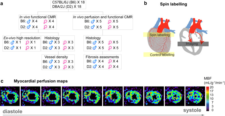
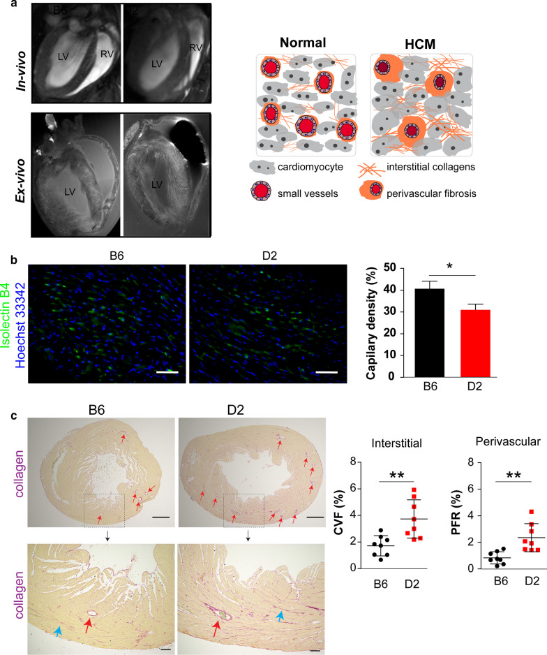
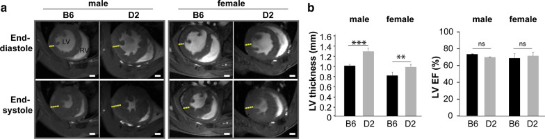
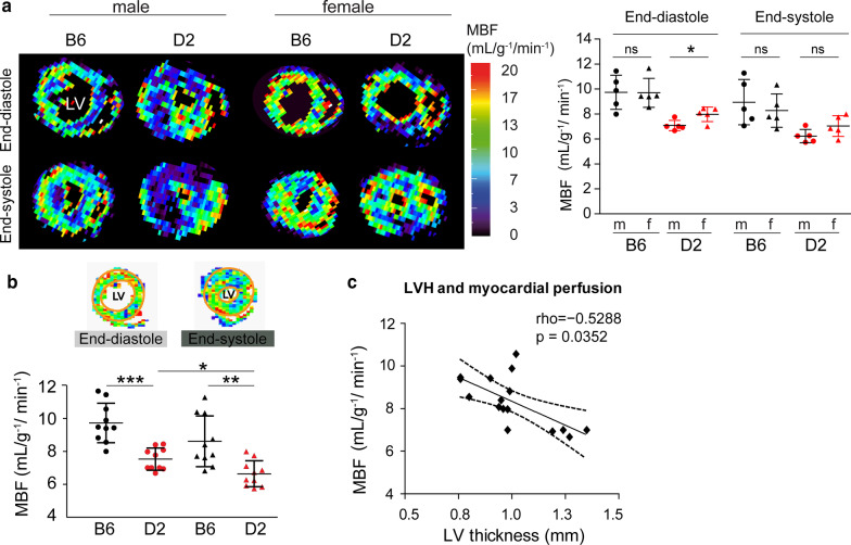
We used a mouse model (DBA/2 J; D2 mouse strain) that spontaneously carries variants in the two most susceptible HCM genes-Mybpc3 and Myh7 and bears the key features of human HCM. The C57BL/6 J (B6) was used as a reference strain. Mice with either B6 or D2 backgrounds (male: n = 4, female: n = 4) underwent cine-CMR for functional assessment at 9.4 T. Left ventricular (LV) wall thickness was measured in end diastolic phase by cine-CMR. Quantitative myocardial perfusion maps (male: n = 5, female: n = 5 in each group) were acquired from arterial spin labelling (cine ASL-CMR) at rest. Myocardial perfusion values were measured by delineating different regions of interest based on the LV segmentation model in the mid ventricle of the LV myocardium. Directly after the CMR, the mouse hearts were removed for histological assessments to confirm the incidence of myocardial interstitial fibrosis (n = 8 in each group) and small vessel remodelling such as vessel density (n = 6 in each group) and perivascular fibrosis (n = 8 in each group).
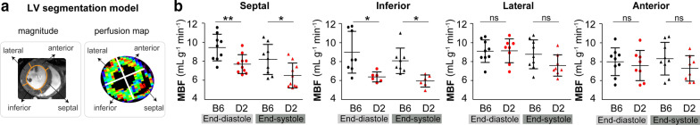
LV hypertrophy was more pronounced in D2 than in B6 mice (male: D2 LV wall thickness = 1.3 ± 0.1 mm vs B6 LV wall thickness = 1.0 ± 0.0 mm, p < 0.001; female: D2 LV wall thickness = 1.0 ± 0.1 mm vs B6 LV wall thickness = 0.8 ± 0.1 mm, p < 0.01). The resting global myocardial perfusion (myocardial blood flow; MBF) was lower in D2 than in B6 mice (end-diastole: D2 MBF = 7.5 ± 0.6 vs B6 MBF = 9.3 ± 1.6 ml/g/min, p < 0.05; end-systole: D2 MBF = 6.6 ± 0.8 vs B6 MBF = 8.2 ± 2.6 ml/g/min, p < 0.01). This myocardial microvascular dysfunction was observed and associated with a reduction in regional MBF, mainly in the interventricular septal and inferior areas of the myocardium. Immunofluorescence revealed a lower number of vessel densities in D2 than in B6 (D2 capillary = 31.0 ± 3.8% vs B6 capillary = 40.7 ± 4.6%, p < 0.05). Myocardial collagen volume fraction (CVF) was significantly higher in D2 LV versus B6 LV mice (D2 CVF = 3.7 ± 1.4% vs B6 CVF = 1.7 ± 0.7%, p < 0.01). Furthermore, a higher ratio of perivascular fibrosis (PFR) was found in D2 than in B6 mice (D2 PFR = 2.3 ± 1.0%, B6 PFR = 0.8 ± 0.4%, p < 0.01).
Our work describes an imaging marker using cine ASL-CMR with a potential to monitor vascular and myocardial remodelling in HCM.
肥厚型心肌病(HCM)相关的心肌血管重塑可能导致心肌血供减少,并随后导致心脏功能逐渐丧失。这一过程很难观察到,因此它们之间的联系仍不清楚。在这里,我们使用非侵入性的心肌血流敏感 CMR 显示了一种在自然发生的 HCM 小鼠模型中静息心肌灌注受损的情况。
我们使用了一种小鼠模型(DBA/2J;D2 小鼠品系),该模型自发携带两种最易患 HCM 的基因-Mybpc3 和 Myh7 的变异体,并具有人类 HCM 的关键特征。C57BL/6J(B6)被用作参考品系。具有 B6 或 D2 背景的雄性(n=4,雌性:n=4)和雌性(n=4)小鼠在 9.4T 下进行电影 CMR 进行功能评估。通过电影 CMR 在舒张末期测量左心室(LV)壁厚度。在休息时从动脉自旋标记(cine ASL-CMR)获得定量心肌灌注图(每组雄性:n=5,雌性:n=5)。通过在 LV 心肌的中心室根据 LV 分段模型勾勒出不同的感兴趣区域来测量心肌灌注值。直接在 CMR 后,取出小鼠心脏进行组织学评估以确认心肌间质纤维化的发生率(每组 n=8)和小血管重塑,如血管密度(每组 n=6)和血管周围纤维化(每组 n=8)。
D2 小鼠的 LV 肥大比 B6 小鼠更为明显(雄性:D2 LV 壁厚度=1.3±0.1mm 与 B6 LV 壁厚度=1.0±0.0mm,p<0.001;雌性:D2 LV 壁厚度=1.0±0.1mm 与 B6 LV 壁厚度=0.8±0.1mm,p<0.01)。与 B6 小鼠相比,D2 小鼠的静息全局心肌灌注(心肌血流;MBF)较低(舒张末期:D2 MBF=7.5±0.6 与 B6 MBF=9.3±1.6ml/g/min,p<0.05;收缩末期:D2 MBF=6.6±0.8 与 B6 MBF=8.2±2.6ml/g/min,p<0.01)。这种心肌微血管功能障碍被观察到,并与区域 MBF 的减少相关,主要是在室间隔和心肌的下区域。免疫荧光显示 D2 小鼠的血管密度低于 B6(D2 毛细血管=31.0±3.8%与 B6 毛细血管=40.7±4.6%,p<0.05)。与 B6 LV 相比,D2 LV 的心肌胶原容积分数(CVF)显著更高(D2 CVF=3.7±1.4%与 B6 CVF=1.7±0.7%,p<0.01)。此外,D2 小鼠的血管周围纤维化(PFR)比率高于 B6 小鼠(D2 PFR=2.3±1.0%与 B6 PFR=0.8±0.4%,p<0.01)。
我们的工作描述了一种使用 cine ASL-CMR 的成像标志物,具有监测 HCM 中血管和心肌重塑的潜力。