Department of Neuroscience and Experimental Therapeutics, Texas A&M University Health Science Center, Bryan, TX, USA.
Department of Neurobiology and Developmental Sciences, University of Arkansas for Medical Sciences, Little Rock, AR, USA.
Alcohol Clin Exp Res. 2021 Jul;45(7):1408-1423. doi: 10.1111/acer.14633. Epub 2021 Jul 1.
The developing hippocampus and cerebellum, unique among brain regions, exhibit a secondary surge in neurogenesis during the third trimester of pregnancy. Ethanol (EtOH) exposure during this period is results in a loss of tissue volume and associated neurobehavioral deficits. However, mechanisms that link EtOH exposure to teratology in these regions are not well understood. We therefore analyzed transcriptomic adaptations to EtOH exposure to identify mechanistic linkages.
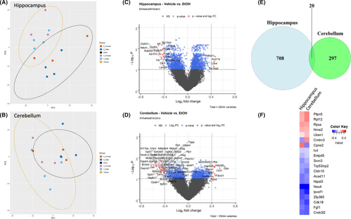
Hippocampi and cerebella were microdissected at postnatal day (P)10, from control C57BL/6J mouse pups, and pups treated with 4 g/kg of EtOH from P4 to P9. RNA was isolated and RNA-seq analysis was performed. We compared gene expression in EtOH- and vehicle-treated control neonates and performed biological pathway-overrepresentation analysis.
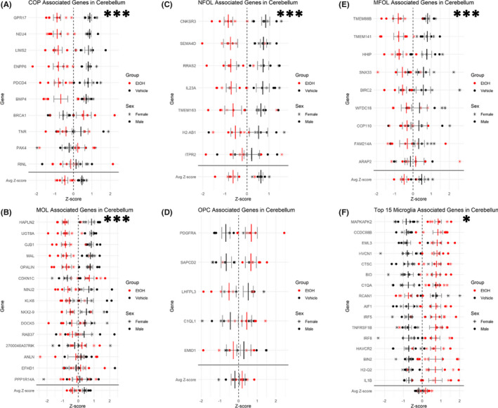
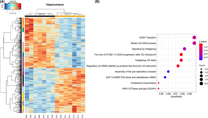
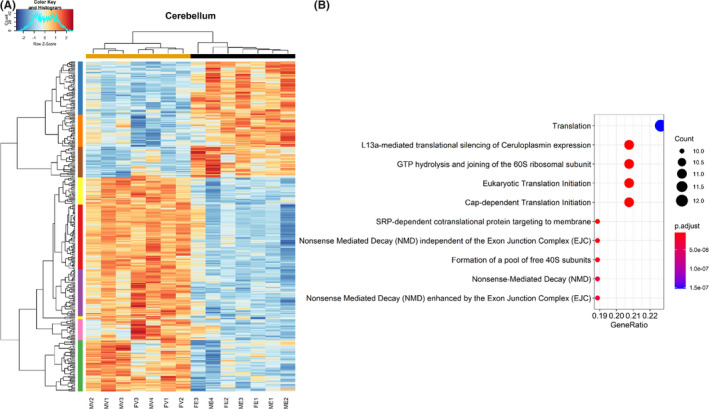
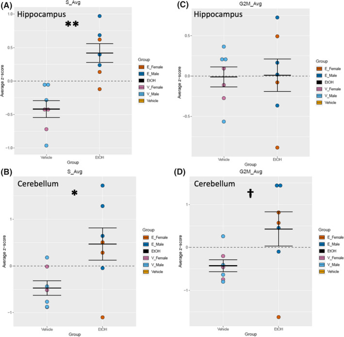
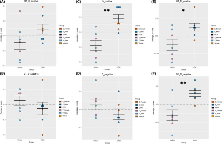
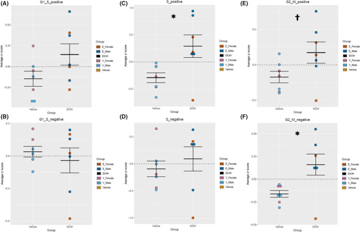
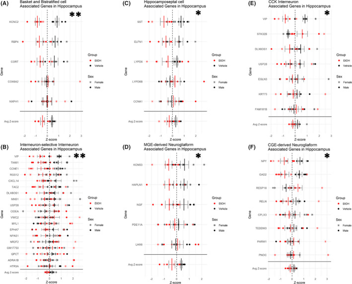
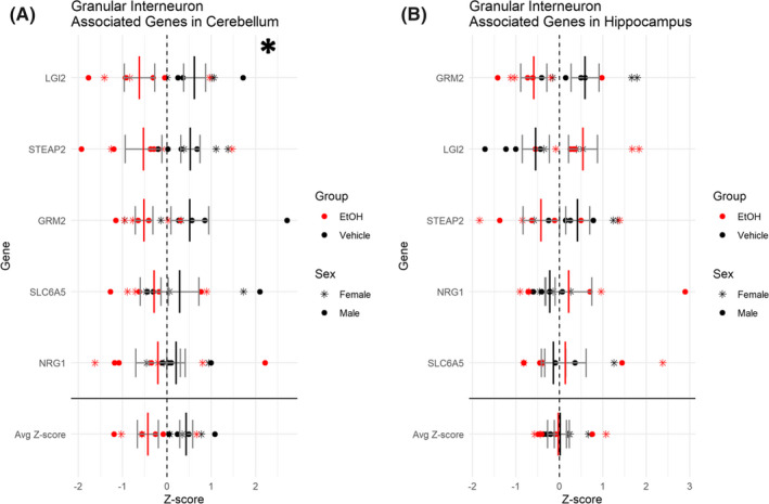
While EtOH exposure resulted in the general induction of genes associated with the S-phase of mitosis in both cerebellum and hippocampus, overall there was little overlap in differentially regulated genes and associated biological pathways between these regions. In cerebellum, EtOH additionally induced gene expression associated with the G2/M-phases of the cell cycle and sonic hedgehog signaling, while in hippocampus, EtOH-induced the pathways for ribosome biogenesis and protein translation. Moreover, EtOH inhibited the transcriptomic identities associated with inhibitory interneuron subpopulations in the hippocampus, while in the cerebellum there was a more pronounced inhibition of transcripts across multiple oligodendrocyte maturation stages.
These data indicate that during the delayed neurogenic period, EtOH may stimulate the cell cycle, but it otherwise results in widely divergent molecular effects in the hippocampus and cerebellum. Moreover, these data provide evidence for region- and cell-type-specific vulnerability, which may contribute to the pathogenic effects of developmental EtOH exposure.
在大脑区域中,发育中的海马体和小脑在妊娠第三个月会出现第二次神经发生激增。在此期间,乙醇(EtOH)暴露会导致组织体积减少和相关的神经行为缺陷。然而,将 EtOH 暴露与这些区域的畸形联系起来的机制尚不清楚。因此,我们分析了转录组适应 EtOH 暴露的情况,以确定潜在的机制联系。
在出生后第 10 天(P10),从对照组 C57BL/6J 幼鼠和从 P4 到 P9 用 4 g/kg EtOH 处理的幼鼠中分离出海马体和小脑。分离出 RNA 并进行 RNA-seq 分析。我们比较了 EtOH 处理和载体处理的对照组新生鼠的基因表达情况,并进行了生物学途径过表达分析。
虽然 EtOH 暴露导致小脑和海马体中与有丝分裂 S 期相关的基因普遍诱导,但这两个区域之间差异调节基因和相关生物学途径的重叠很少。在小脑体中,EtOH 还诱导了与细胞周期 G2/M 期和 Sonic Hedgehog 信号相关的基因表达,而在海马体中,EtOH 诱导了核糖体生物发生和蛋白质翻译的途径。此外,EtOH 抑制了海马体中抑制性中间神经元亚群的转录本特征,而在小脑体中,多个少突胶质细胞成熟阶段的转录本抑制更为明显。
这些数据表明,在延迟的神经发生期,EtOH 可能刺激细胞周期,但在海马体和小脑体中会导致广泛不同的分子效应。此外,这些数据为区域和细胞类型特异性易感性提供了证据,这可能导致发育性 EtOH 暴露的致病效应。