Fernández-Espejo Emilio, Rodríguez de Fonseca Fernando, Suárez Juan, Tolosa Eduardo, Vilas Dolores, Aldecoa Iban, Berenguer Joan, Damas-Hermoso Fátima
Reial Acadèmia de Medicina de Catalunya, 08001 Barcelona, Spain.
Red Andaluza de Investigación Clínica y Traslacional en Neurología (Neuro-RECA), Laboratorio de Medicina Regenerativa, Hospital Regional Universitario, 29010 Málaga, Spain.
Antioxidants (Basel). 2021 May 1;10(5):715. doi: 10.3390/antiox10050715.
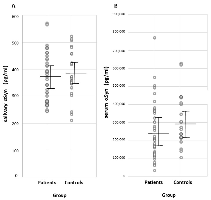
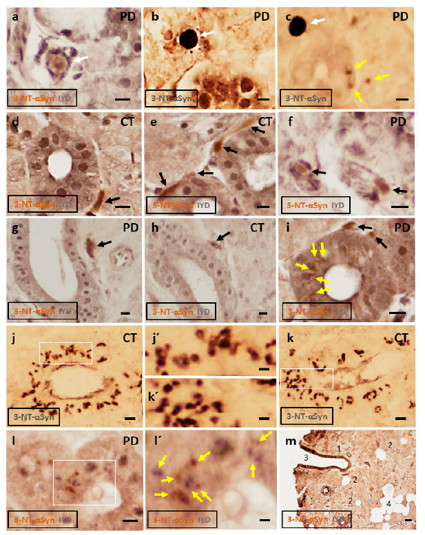
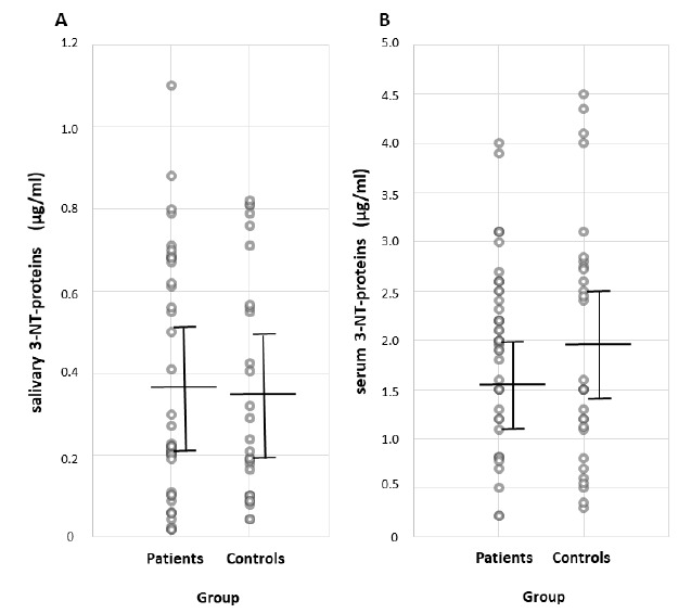
Salivary α-synuclein (aSyn) and its nitrated form, or 3-nitrotyrosine-α-synuclein (3-NT-αSyn), hold promise as biomarkers for idiopathic Parkinson's disease (IPD). Nitrative stress that is characterized by an excess of 3-nitrotyrosine proteins (3-NT-proteins) has been proposed as a pathogenic mechanism in IPD. The objective is to study the pathological role of native αSyn, 3-NT-αSyn, and 3-NT-proteins in the saliva and submandibulary glands of patients with IPD. The salivary and serum αSyn and 3-NT-proteins concentration is evaluated with ELISA in patients and controls. Correlations of αSyn and 3-NT-proteins content with clinical features of the disease are examined. Immunohistochemical 3-NT-αSyn expression in submandibulary gland sections is analyzed. (a) Salivary concentration and saliva/serum ratios of native αSyn and 3-NT-proteins are similar in patients and controls; (b) salivary αSyn and 3-NT-proteins do not correlate with any clinical feature; and (c) three patterns of 3-NT-αSyn-positive inclusions are observed on histological sections: rounded "Lewy-type" aggregates of 10-25 µm in diameter, coarse deposits with varied morphology, and spheroid inclusions or bodies of 3-5 µm in diameter. "Lewy-type" and coarse inclusions are observed in the interlobular connective tissue of the gland, and small-sized bodies are located within the cytoplasm of duct cells. "Lewy-type" inclusions are only observed in patients, and the remaining patterns of inclusions are observed in both the patients and controls. The patients' saliva presents a similar concentration of native αSyn and 3-nitrotyrosine-proteins than that of the controls, and no correlations with clinical features are found. These findings preclude the utility of native αSyn in the saliva as a biomarker, and they indicate the absence of nitrative stress in the saliva and serum of patients. As regards nitrated αSyn, "Lewy-type" inclusions expressing 3-NT-αSyn are observed in the patients, not the controls-a novel finding that suggests that a biopsy of the submandibulary gland, if proven safe, could be a useful technique for diagnosing IPD. Finally, to our knowledge, this is also the first description of 3-NT-αSyn-immunoreactive intracytoplasmic bodies in cells that are located outside the nervous system. These intracytoplasmic bodies are present in duct cells of submandibulary gland sections from all subjects regardless of their pathology, and they can represent an aging or involutional change. Further immunostaining studies with different antibodies and larger samples are needed to validate the data.
唾液α-突触核蛋白(aSyn)及其硝化形式,即3-硝基酪氨酸-α-突触核蛋白(3-NT-αSyn),有望成为特发性帕金森病(IPD)的生物标志物。以过量的3-硝基酪氨酸蛋白(3-NT-蛋白)为特征的硝化应激已被提出是IPD的一种致病机制。目的是研究天然αSyn、3-NT-αSyn和3-NT-蛋白在IPD患者唾液和下颌下腺中的病理作用。通过酶联免疫吸附测定(ELISA)评估患者和对照组唾液及血清中的αSyn和3-NT-蛋白浓度。检查αSyn和3-NT-蛋白含量与疾病临床特征的相关性。分析下颌下腺切片中3-NT-αSyn的免疫组化表达。(a)患者和对照组中天然αSyn和3-NT-蛋白的唾液浓度及唾液/血清比值相似;(b)唾液αSyn和3-NT-蛋白与任何临床特征均无相关性;(c)在组织学切片上观察到三种3-NT-αSyn阳性包涵体模式:直径为10 - 25μm的圆形“路易小体型”聚集体、形态各异的粗大沉积物以及直径为3 - 5μm的球形包涵体或小体。“路易小体型”和粗大包涵体见于腺体的小叶间结缔组织,小体位于导管细胞的细胞质内。“路易小体型”仅在患者中观察到,其余包涵体模式在患者和对照组中均有观察到。患者唾液中天然αSyn和3-硝基酪氨酸蛋白的浓度与对照组相似,且未发现与临床特征相关。这些发现排除了唾液中天然αSyn作为生物标志物的效用,并表明患者唾液和血清中不存在硝化应激。关于硝化αSyn,在患者而非对照组中观察到表达3-NT-αSyn的“路易小体型”包涵体——这一新颖发现表明,如果经证实安全,下颌下腺活检可能是诊断IPD的一种有用技术。最后,据我们所知,这也是对位于神经系统外的细胞中3-NT-αSyn免疫反应性胞质小体的首次描述。这些胞质小体存在于所有受试者下颌下腺切片的导管细胞中,无论其病理情况如何,它们可能代表一种衰老或退化性变化。需要用不同抗体和更大样本进行进一步的免疫染色研究以验证数据。