Department of Ophthalmology, Medical University of South Carolina, Charleston, SC 29425, USA.
Ralph H. Johnson VA Medical Center, Division of Research, Charleston, SC 29401, USA.
Int J Mol Sci. 2021 May 11;22(10):5083. doi: 10.3390/ijms22105083.
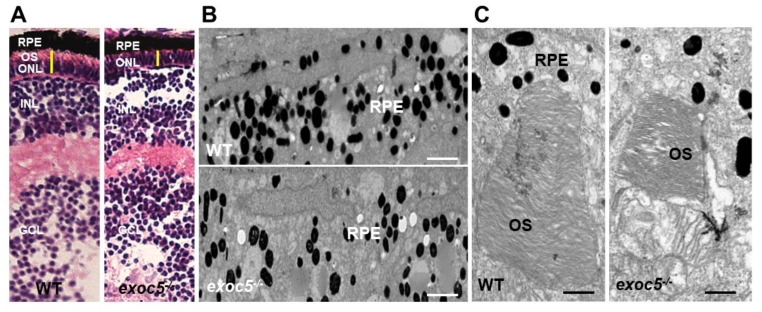
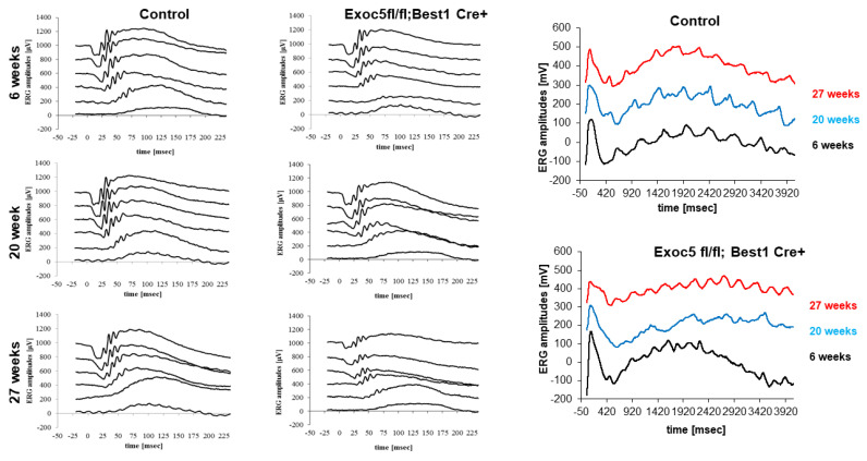
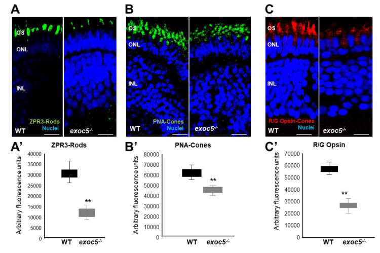
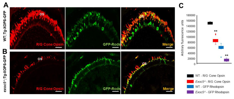
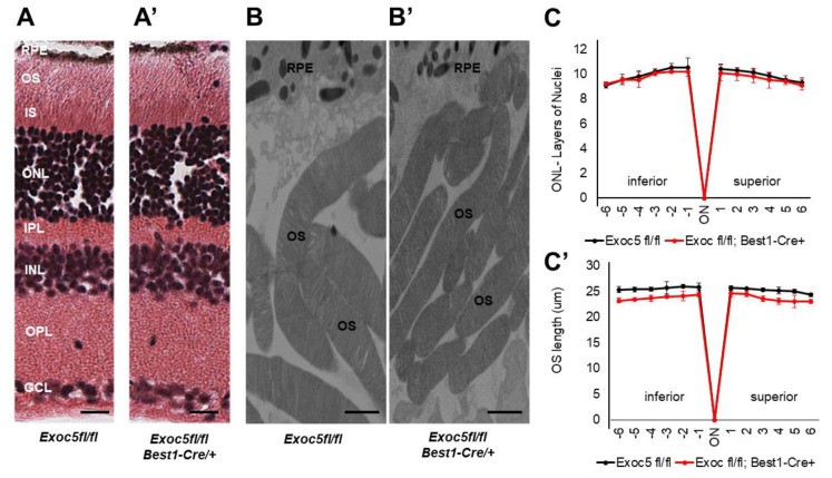
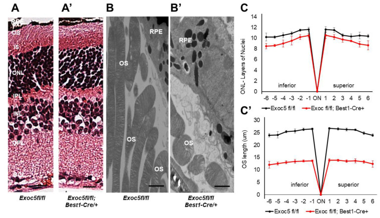
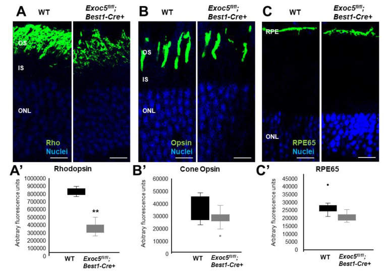
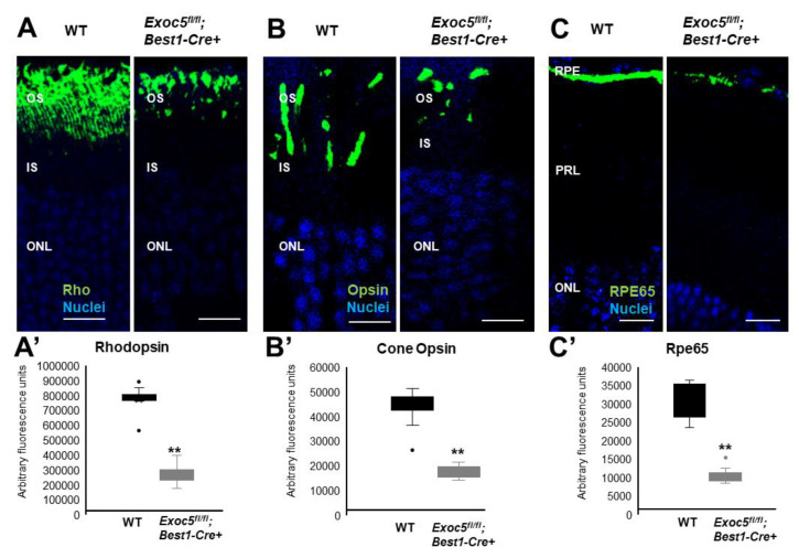
To characterize the mechanisms by which the highly conserved exocyst trafficking complex regulates eye physiology in zebrafish and mice, we focused on Exoc5 (also known as ), a central exocyst component. We analyzed both zebrafish mutants and retinal pigmented epithelium (RPE)-specific knockout mice. Exoc5 is present in both the non-pigmented epithelium of the ciliary body and in the RPE. In this study, we set out to establish an animal model to study the mechanisms underlying the ocular phenotype and to establish if loss of visual function is induced by postnatal RPE Exoc5-deficiency. zebrafish had smaller eyes, with decreased number of melanocytes in the RPE and shorter photoreceptor outer segments. At 3.5 days post-fertilization, loss of rod and cone opsins were observed in zebrafish mutants. Mice with postnatal RPE-specific loss of Exoc5 showed retinal thinning associated with compromised visual function and loss of visual photoreceptor pigments. Abnormal levels of RPE65 together with a reduced c-wave amplitude indicate a dysfunctional RPE. The retinal phenotype in mice was present at 20 weeks, but was more pronounced at 27 weeks, indicating progressive disease phenotype. We previously showed that the exocyst is necessary for photoreceptor ciliogenesis and retinal development. Here, we report that mutant zebrafish and mice with RPE-specific genetic ablation of Exoc5 develop abnormal RPE pigmentation, resulting in retinal cell dystrophy and loss of visual pigments associated with compromised vision. Together, these data suggest that exocyst-mediated signaling in the RPE is required for RPE structure and function, indirectly leading to photoreceptor degeneration.
为了阐明高度保守的外泌体运输复合物在调控斑马鱼和小鼠眼睛生理中的作用机制,我们聚焦于外泌体复合物的一个核心组分 Exoc5(也称为 )。我们分析了 突变的斑马鱼和视网膜色素上皮(RPE)特异性 敲除的小鼠。Exoc5 存在于睫状体的非色素上皮和 RPE 中。在本研究中,我们旨在建立一个动物模型来研究眼部表型的潜在机制,并确定 RPE 中 Exoc5 缺失是否会导致视觉功能丧失。 突变的斑马鱼眼睛较小,RPE 中的黑色素细胞数量减少,光感受器外节变短。在受精后 3.5 天,我们观察到 突变的斑马鱼中杆状和锥状视蛋白缺失。在出生后 RPE 特异性缺失 Exoc5 的小鼠中,视网膜变薄与视觉功能受损和视觉光感受器色素丧失有关。RPE65 水平异常和 c 波幅度降低表明 RPE 功能障碍。在 小鼠中,视网膜表型在 20 周时已经存在,但在 27 周时更为明显,表明疾病呈进行性发展。我们之前表明,外泌体对于光感受器纤毛发生和视网膜发育是必需的。在这里,我们报告 突变的斑马鱼和 RPE 特异性遗传消融 Exoc5 的小鼠会出现异常的 RPE 色素沉着,导致视网膜细胞变性和与视觉功能受损相关的视觉色素丧失。总之,这些数据表明,RPE 中的外泌体介导的信号对于 RPE 的结构和功能是必需的,这间接导致了光感受器的退化。