• 文献检索
  • 文档翻译
  • 深度研究
  • 学术资讯
  • Suppr Zotero 插件Zotero 插件
  • 邀请有礼
  • 套餐&价格
  • 历史记录
应用&插件
Suppr Zotero 插件Zotero 插件浏览器插件Mac 客户端Windows 客户端微信小程序
定价
高级版会员购买积分包购买API积分包
服务
文献检索文档翻译深度研究API 文档MCP 服务
关于我们
关于 Suppr公司介绍联系我们用户协议隐私条款
关注我们

Suppr 超能文献

核心技术专利:CN118964589B侵权必究
粤ICP备2023148730 号-1Suppr @ 2026

文献检索

告别复杂PubMed语法,用中文像聊天一样搜索,搜遍4000万医学文献。AI智能推荐,让科研检索更轻松。

立即免费搜索

文件翻译

保留排版,准确专业,支持PDF/Word/PPT等文件格式,支持 12+语言互译。

免费翻译文档

深度研究

AI帮你快速写综述,25分钟生成高质量综述,智能提取关键信息,辅助科研写作。

立即免费体验

双相情感障碍缓解期静息态脑的时频特征——一项初步研究

Time-Frequency Characterization of Resting Brain in Bipolar Disorder during Euthymia-A Preliminary Study.

作者信息

Chrobak Adrian Andrzej, Bohaterewicz Bartosz, Sobczak Anna Maria, Marszał-Wiśniewska Magdalena, Tereszko Anna, Krupa Anna, Ceglarek Anna, Fafrowicz Magdalena, Bryll Amira, Marek Tadeusz, Dudek Dominika, Siwek Marcin

机构信息

Department of Adult Psychiatry, Jagiellonian University Medical College, Kopernika st. 21a, 31-501 Kraków, Poland.

Department of Psychology of Individual Differences, Psychological Diagnosis and Psychometrics, Faculty of Psychology in Warsaw, SWPS University of Social Sciences and Humanities, Chodakowska st. 19/31, 03-815 Warsaw, Poland.

出版信息

Brain Sci. 2021 May 7;11(5):599. doi: 10.3390/brainsci11050599.

DOI:10.3390/brainsci11050599
PMID:34067189
原文链接:https://pmc.ncbi.nlm.nih.gov/articles/PMC8150994/
Abstract

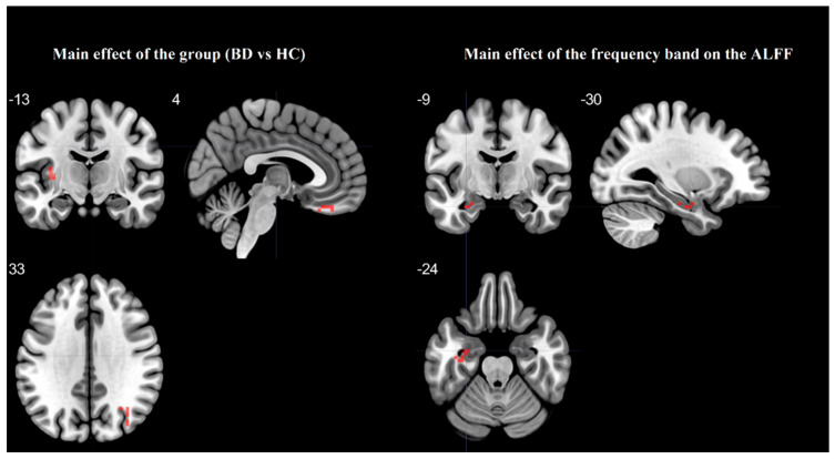
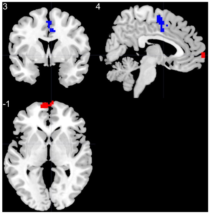
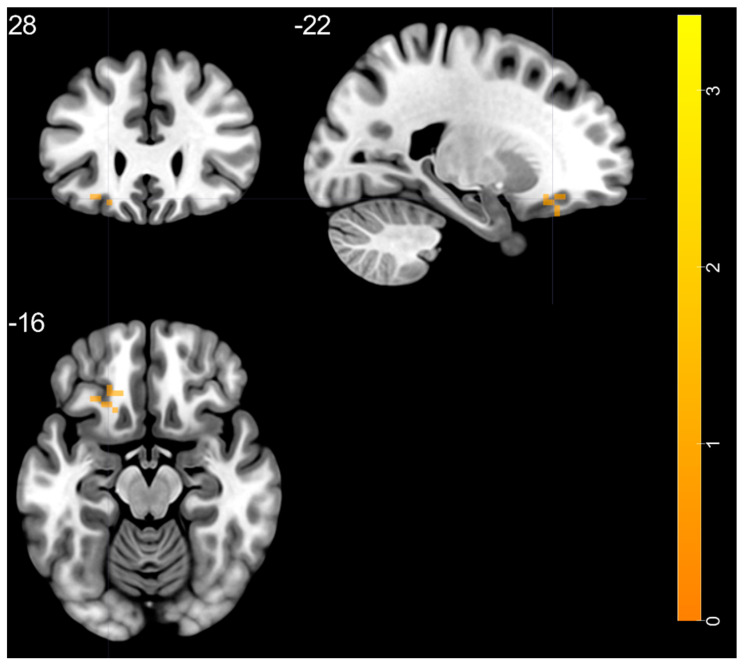
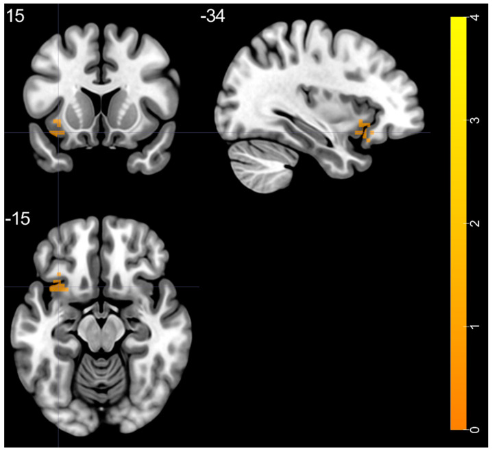
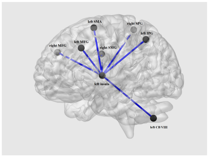
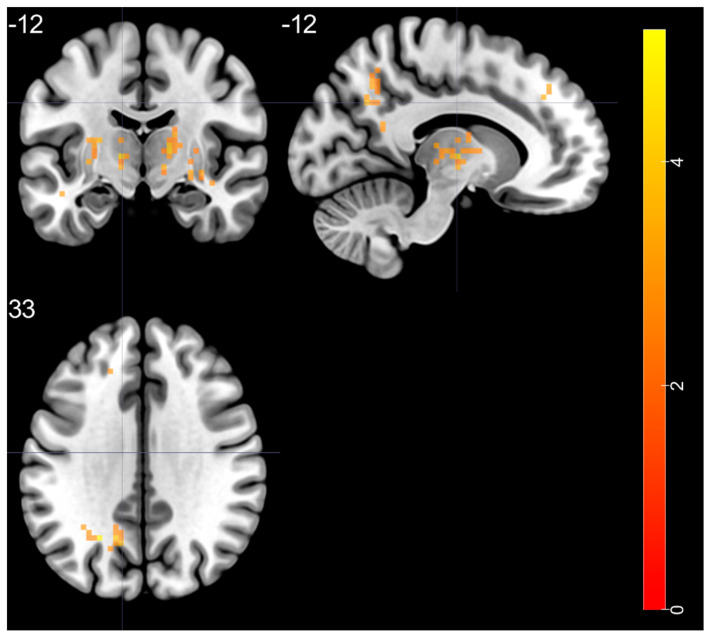
The goal of this paper is to investigate the baseline brain activity in euthymic bipolar disorder (BD) patients by comparing it to healthy controls (HC) with the use of a variety of resting state functional magnetic resonance imaging (rs-fMRI) analyses, such as amplitude of low frequency fluctuations (ALFF), fractional ALFF (f/ALFF), ALFF-based functional connectivity (FC), and r egional homogeneity (ReHo). We hypothesize that above-mentioned techniques will differentiate BD from HC indicating dissimilarities between the groups within different brain structures. Forty-two participants divided into two groups of euthymic BD patients (n = 21) and HC (n = 21) underwent rs-fMRI evaluation. Typical band ALFF, slow-4, slow-5, f/ALFF, as well as ReHo indexes were analyzed. Regions with altered ALFF were chosen as ROI for seed-to-voxel analysis of FC. As opposed to HC, BD patients revealed: increased ALFF in left insula; increased slow-5 in left middle temporal pole; increased f/ALFF in left superior frontal gyrus, left superior temporal gyrus, left middle occipital gyrus, right putamen, and bilateral thalamus. There were no significant differences between BD and HC groups in slow-4 band. Compared to HC, the BD group presented higher ReHo values in the left superior medial frontal gyrus and lower ReHo values in the right supplementary motor area. FC analysis revealed significant hyper-connectivity within the BD group between left insula and bilateral middle frontal gyrus, right superior parietal gyrus, right supramarginal gyrus, left inferior parietal gyrus, left cerebellum, and left supplementary motor area. To our best knowledge, this is the first rs-fMRI study combining ReHo, ALFF, f/ALFF, and subdivided frequency bands (slow-4 and slow-5) in euthymic BD patients. ALFF, f/ALFF, slow-5, as well as REHO analysis revealed significant differences between two studied groups. Although results obtained with the above methods enable to identify group-specific brain structures, no overlap between the brain regions was detected. This indicates that combination of foregoing rs-fMRI methods may complement each other, revealing the bigger picture of the complex resting state abnormalities in BD.

摘要

本文的目的是通过使用多种静息态功能磁共振成像(rs-fMRI)分析方法,如低频波动幅度(ALFF)、分数ALFF(f/ALFF)、基于ALFF的功能连接(FC)和局部一致性(ReHo),将双相情感障碍(BD)缓解期患者的基线脑活动与健康对照(HC)进行比较,以此来研究BD缓解期患者的基线脑活动。我们假设上述技术能够区分BD患者和HC,表明不同脑结构组之间存在差异。42名参与者被分为两组,即BD缓解期患者组(n = 21)和HC组(n = 21),并接受了rs-fMRI评估。分析了典型频段的ALFF、慢波4、慢波5、f/ALFF以及ReHo指数。将ALFF改变的区域选为感兴趣区(ROI),用于FC的种子点到体素分析。与HC相比,BD患者表现出:左侧岛叶的ALFF增加;左侧颞中极的慢波5增加;左侧额上回、左侧颞上回、左侧枕中回、右侧壳核和双侧丘脑的f/ALFF增加。BD组和HC组在慢波4频段没有显著差异。与HC相比,BD组在左侧额内侧上回的ReHo值较高,而在右侧辅助运动区的ReHo值较低。FC分析显示,BD组内左侧岛叶与双侧额中回、右侧顶上小叶、右侧缘上回、左侧顶下小叶、左侧小脑和左侧辅助运动区之间存在显著的高连接性。据我们所知,这是第一项在BD缓解期患者中结合ReHo、ALFF、f/ALFF和细分频段(慢波4和慢波5)的rs-fMRI研究。ALFF、f/ALFF、慢波5以及ReHo分析显示,两个研究组之间存在显著差异。尽管上述方法获得的结果能够识别特定组的脑结构,但未检测到脑区之间的重叠。这表明上述rs-fMRI方法的组合可能会相互补充,揭示BD复杂静息态异常的全貌。

https://cdn.ncbi.nlm.nih.gov/pmc/blobs/6607/8150994/0baa51257508/brainsci-11-00599-g006.jpg
https://cdn.ncbi.nlm.nih.gov/pmc/blobs/6607/8150994/63166599a8b3/brainsci-11-00599-g001.jpg
https://cdn.ncbi.nlm.nih.gov/pmc/blobs/6607/8150994/1e6ba8f22db0/brainsci-11-00599-g002.jpg
https://cdn.ncbi.nlm.nih.gov/pmc/blobs/6607/8150994/01fcb1bd4230/brainsci-11-00599-g003.jpg
https://cdn.ncbi.nlm.nih.gov/pmc/blobs/6607/8150994/19ba636767a1/brainsci-11-00599-g004.jpg
https://cdn.ncbi.nlm.nih.gov/pmc/blobs/6607/8150994/467881cecfc8/brainsci-11-00599-g005.jpg
https://cdn.ncbi.nlm.nih.gov/pmc/blobs/6607/8150994/0baa51257508/brainsci-11-00599-g006.jpg

相似文献

1
Time-Frequency Characterization of Resting Brain in Bipolar Disorder during Euthymia-A Preliminary Study.双相情感障碍缓解期静息态脑的时频特征——一项初步研究
Brain Sci. 2021 May 7;11(5):599. doi: 10.3390/brainsci11050599.
2
Comparison of resting-state spontaneous brain activity between treatment-naive schizophrenia and obsessive-compulsive disorder.治疗初发精神分裂症与强迫症患者静息态自发脑活动的比较。
BMC Psychiatry. 2021 Nov 3;21(1):544. doi: 10.1186/s12888-021-03554-y.
3
Frequency-Dependent Alterations in the Amplitude of Low-Frequency Fluctuations in Patients with Acute Pericoronitis: A Resting-State fMRI Study.急性智齿冠周炎患者低频波动振幅的频率依赖性改变:一项静息态功能磁共振成像研究
J Pain Res. 2023 Feb 15;16:501-511. doi: 10.2147/JPR.S397523. eCollection 2023.
4
Common and distinct patterns of intrinsic brain activity alterations in major depression and bipolar disorder: voxel-based meta-analysis.基于体素的荟萃分析:重度抑郁症和双相情感障碍患者大脑固有活动改变的常见和独特模式。
Transl Psychiatry. 2020 Oct 19;10(1):353. doi: 10.1038/s41398-020-01036-5.
5
Aberrant amplitude low-frequency fluctuation (ALFF) and regional homogeneity (ReHo) in generalized anxiety disorder (GAD) and their roles in predicting treatment remission.广泛性焦虑障碍(GAD)中的异常振幅低频波动(ALFF)和局部一致性(ReHo)及其在预测治疗缓解中的作用。
Ann Transl Med. 2020 Oct;8(20):1319. doi: 10.21037/atm-20-6448.
6
Altered Brain Function in Treatment-Resistant and Non-treatment-resistant Depression Patients: A Resting-State Functional Magnetic Resonance Imaging Study.难治性和非难治性抑郁症患者的脑功能改变:一项静息态功能磁共振成像研究
Front Psychiatry. 2022 Jul 22;13:904139. doi: 10.3389/fpsyt.2022.904139. eCollection 2022.
7
The rs-fMRI study of effects of fornix and hippocampus-related brain function after the transcallosal interforniceal approach.经胼胝体间穹窿入路切除后穹窿和海马相关脑区的功能改变的 rs-fMRI 研究。
Brain Res Bull. 2019 Aug;150:207-215. doi: 10.1016/j.brainresbull.2019.05.014. Epub 2019 May 22.
8
Resting-state fMRI signals in offspring of parents with bipolar disorder at the high-risk and ultra-high-risk stages and their relations with cognitive function.处于高危和超高危阶段的双相障碍父母的后代的静息态 fMRI 信号及其与认知功能的关系。
J Psychiatr Res. 2018 Mar;98:99-106. doi: 10.1016/j.jpsychires.2018.01.001. Epub 2018 Jan 8.
9
Alterations of spontaneous brain activity in systematic lupus erythematosus patients without neuropsychiatric symptoms: A resting-functional MRI study.无神经精神症状的系统性红斑狼疮患者自发脑活动的改变:一项静息态功能磁共振成像研究
Lupus. 2021 Oct;30(11):1781-1789. doi: 10.1177/09612033211033984. Epub 2021 Oct 8.
10
Aberrant Spontaneous Brain Activity and its Association with Cognitive Function in Non-Obese Nonalcoholic Fatty Liver Disease: A Resting-State fMRI Study.非肥胖非酒精性脂肪性肝病患者的自发性脑活动异常及其与认知功能的关系:一项静息态 fMRI 研究。
J Integr Neurosci. 2023 Jan 4;22(1):8. doi: 10.31083/j.jin2201008.

引用本文的文献

1
Altered brain regional homogeneity, depressive symptoms, and cognitive impairments in medication-free female patients with current depressive episodes in bipolar disorder and major depressive disorder.双相情感障碍和重度抑郁症中目前有抑郁发作的未服药女性患者的脑区同质性改变、抑郁症状及认知障碍。
BMC Psychiatry. 2024 Dec 6;24(1):892. doi: 10.1186/s12888-024-06352-4.
2
Divergent pattern of functional connectivity within the dorsal attention network differentiates schizophrenia and bipolar disorder patients.背侧注意网络内功能连接的不同模式可区分精神分裂症患者和双相情感障碍患者。
Front Psychiatry. 2024 Sep 19;15:1474313. doi: 10.3389/fpsyt.2024.1474313. eCollection 2024.
3

本文引用的文献

1
Altered Functional Connectivity Differences in Salience Network as a Neuromarker of Suicide Risk in Euthymic Bipolar Disorder Patients.心境正常的双相情感障碍患者中,突显网络功能连接差异的改变作为自杀风险的神经标志物
Front Hum Neurosci. 2020 Nov 13;14:585766. doi: 10.3389/fnhum.2020.585766. eCollection 2020.
2
Altered functional connectivity among frontal eye fields, thalamus and cerebellum in bipolar disorder.双相情感障碍患者额叶眼区、丘脑和小脑之间的功能连接改变。
Psychiatr Pol. 2019 Jun 30;54(3):487-497. doi: 10.12740/PP/OnlineFirst/104445. Epub 2020 Jun 30.
3
Surface-based regional homogeneity in bipolar disorder: A resting-state fMRI study.
Decreased dynamic variability of the cerebellum in the euthymic patients with bipolar disorder.
双相情感障碍缓解期患者小脑动态变异性降低。
BMC Psychiatry. 2024 Feb 19;24(1):137. doi: 10.1186/s12888-024-05596-4.
4
When practice does not make a perfect - paradoxical learning curve in schizophrenia and bipolar disorder revealed by different serial reaction time task variants.当练习无法造就完美——精神分裂症和双相情感障碍中不同序列反应时任务变体所揭示的矛盾学习曲线。
Front Psychiatry. 2023 Sep 12;14:1238473. doi: 10.3389/fpsyt.2023.1238473. eCollection 2023.
5
The atypical functional connectivity of Broca's area at multiple frequency bands in autism spectrum disorder.自闭症谱系障碍中布罗卡区在多个频段的非典型功能连接。
Brain Imaging Behav. 2022 Dec;16(6):2627-2636. doi: 10.1007/s11682-022-00718-6. Epub 2022 Sep 26.
6
Altered Brain Function in Treatment-Resistant and Non-treatment-resistant Depression Patients: A Resting-State Functional Magnetic Resonance Imaging Study.难治性和非难治性抑郁症患者的脑功能改变:一项静息态功能磁共振成像研究
Front Psychiatry. 2022 Jul 22;13:904139. doi: 10.3389/fpsyt.2022.904139. eCollection 2022.
7
Alterations in resting-state whole-brain functional connectivity pattern similarity in bipolar disorder patients.双相障碍患者静息态全脑功能连接模式相似性的改变。
Brain Behav. 2022 May;12(5):e2580. doi: 10.1002/brb3.2580. Epub 2022 Apr 21.
8
Neural Networks and Connectivity among Brain Regions.神经网络与脑区之间的连接性
Brain Sci. 2022 Mar 3;12(3):346. doi: 10.3390/brainsci12030346.
基于体素的脑区局部一致性分析在双相障碍中的研究:一项静息态 fMRI 研究。
Psychiatry Res. 2019 Aug;278:199-204. doi: 10.1016/j.psychres.2019.05.045. Epub 2019 May 31.
4
Altered regional homogeneity in pediatric bipolar disorder during manic and euthymic state: a resting-state fMRI study.儿童双相障碍在躁狂和缓解期的局部一致性改变:一项静息态 fMRI 研究。
Brain Imaging Behav. 2019 Dec;13(6):1789-1798. doi: 10.1007/s11682-019-00117-4.
5
Spontaneous low-frequency fluctuations in the neural system for emotional perception in major psychiatric disorders: amplitude similarities and differences across frequency bands.重大精神障碍情绪感知的神经系统自发性低频波动:各频段的幅度相似性和差异性。
J Psychiatry Neurosci. 2019 Mar 1;44(2):132-141. doi: 10.1503/jpn.170226.
6
Resting state connectivity differences in eyes open versus eyes closed conditions.睁眼与闭眼状态下的静息态连接差异。
Hum Brain Mapp. 2019 Jun 1;40(8):2488-2498. doi: 10.1002/hbm.24539. Epub 2019 Feb 5.
7
Neural signatures of the risk for bipolar disorder: A meta-analysis of structural and functional neuroimaging studies.双相障碍风险的神经特征:结构和功能神经影像学研究的荟萃分析。
Bipolar Disord. 2019 May;21(3):215-227. doi: 10.1111/bdi.12720. Epub 2018 Dec 14.
8
The Abnormality of Topological Asymmetry in Hemispheric Brain Anatomical Networks in Bipolar Disorder.双相情感障碍患者大脑半球解剖网络中拓扑不对称性的异常
Front Neurosci. 2018 Sep 3;12:618. doi: 10.3389/fnins.2018.00618. eCollection 2018.
9
Resting-state functional connectivity in individuals with bipolar disorder during clinical remission: a systematic review.双相障碍患者临床缓解期静息态功能连接的系统评价。
J Psychiatry Neurosci. 2018 Aug;43(5):298-316. doi: 10.1503/jpn.170175.
10
Neural Basis of Depression Related to a Dominant Right Hemisphere: A Resting-State fMRI Study.与右半球优势相关的抑郁症的神经基础:一项静息态功能磁共振成像研究。
Behav Neurol. 2018 Jun 5;2018:5024520. doi: 10.1155/2018/5024520. eCollection 2018.