Department of Psychology, University of South Carolina, Columbia, SC 29208, USA.
Viruses. 2021 May 17;13(5):924. doi: 10.3390/v13050924.
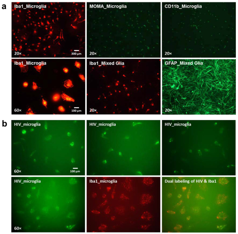
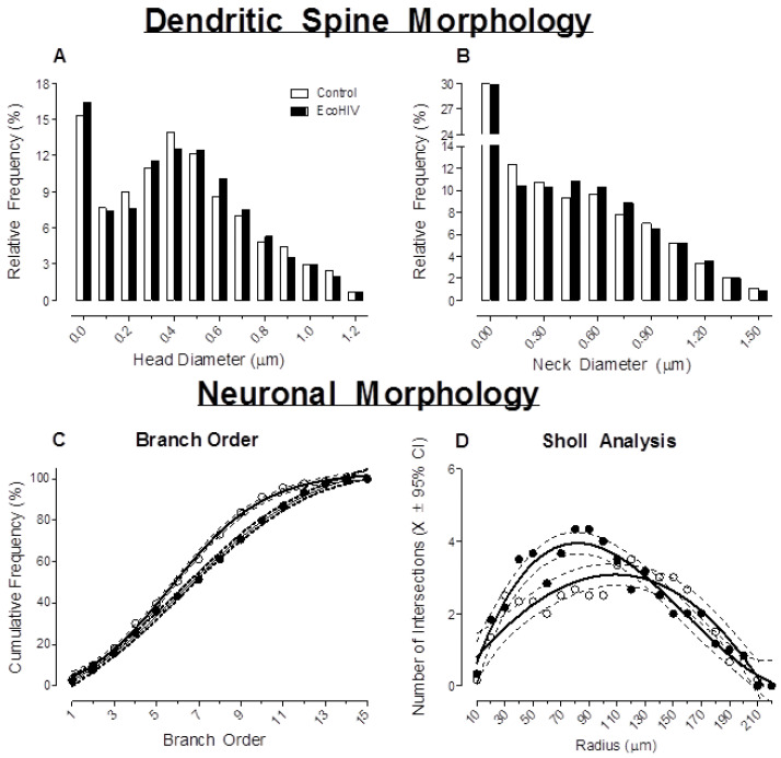
The persistence of HIV-1 viral reservoirs in the brain, despite treatment with combination antiretroviral therapy (cART), remains a critical roadblock for the development of a novel cure strategy for HIV-1. To enhance our understanding of viral reservoirs, two complementary studies were conducted to (1) evaluate the HIV-1 mRNA distribution pattern and major cell type expressing HIV-1 mRNA in the HIV-1 transgenic (Tg) rat, and (2) validate our findings by developing and critically testing a novel biological system to model active HIV-1 infection in the rat. First, a restricted, region-specific HIV-1 mRNA distribution pattern was observed in the HIV-1 Tg rat. Microglia were the predominant cell type expressing HIV-1 mRNA in the HIV-1 Tg rat. Second, we developed and critically tested a novel biological system to model key aspects of HIV-1 by infusing F344/N control rats with chimeric HIV (EcoHIV). In vitro, primary cultured microglia were treated with EcoHIV revealing prominent expression within 24 h of infection. In vivo, EcoHIV expression was observed seven days after stereotaxic injections. Following EcoHIV infection, microglia were the major cell type expressing HIV-1 mRNA, results that are consistent with observations in the HIV-1 Tg rat. Within eight weeks of infection, EcoHIV rats exhibited neurocognitive impairments and synaptic dysfunction, which may result from activation of the NogoA-NgR3/PirB-RhoA signaling pathway and/or neuroinflammation. Collectively, these studies enhance our understanding of HIV-1 viral reservoirs in the brain and offer a novel biological system to model HIV-associated neurocognitive disorders and associated comorbidities (i.e., drug abuse) in rats.
尽管采用联合抗逆转录病毒疗法 (cART) 治疗,但 HIV-1 病毒库在大脑中的持续存在仍然是开发新型 HIV-1 治疗策略的关键障碍。为了增强我们对病毒库的理解,进行了两项互补研究:(1)评估 HIV-1 转基因 (Tg) 大鼠中 HIV-1 mRNA 的分布模式和主要表达 HIV-1 mRNA 的细胞类型;(2)通过开发和严格测试一种新的生物系统来模拟大鼠中的活跃 HIV-1 感染,验证我们的发现。首先,在 HIV-1 Tg 大鼠中观察到受限的、特定区域的 HIV-1 mRNA 分布模式。小胶质细胞是 HIV-1 Tg 大鼠中表达 HIV-1 mRNA 的主要细胞类型。其次,我们开发并严格测试了一种新的生物系统,通过向 F344/N 对照大鼠输注嵌合 HIV (EcoHIV) 来模拟 HIV-1 的关键方面。在体外,用 EcoHIV 处理原代培养的小胶质细胞,在感染后 24 小时内即可观察到明显的表达。在体内,在立体定向注射后 7 天观察到 EcoHIV 的表达。EcoHIV 感染后,小胶质细胞是表达 HIV-1 mRNA 的主要细胞类型,这些结果与 HIV-1 Tg 大鼠的观察结果一致。感染后 8 周,EcoHIV 大鼠表现出神经认知障碍和突触功能障碍,这可能是由于 NogoA-NgR3/PirB-RhoA 信号通路的激活和/或神经炎症所致。总的来说,这些研究增强了我们对大脑中 HIV-1 病毒库的理解,并提供了一种新的生物系统来模拟大鼠中的 HIV 相关神经认知障碍和相关合并症(即药物滥用)。